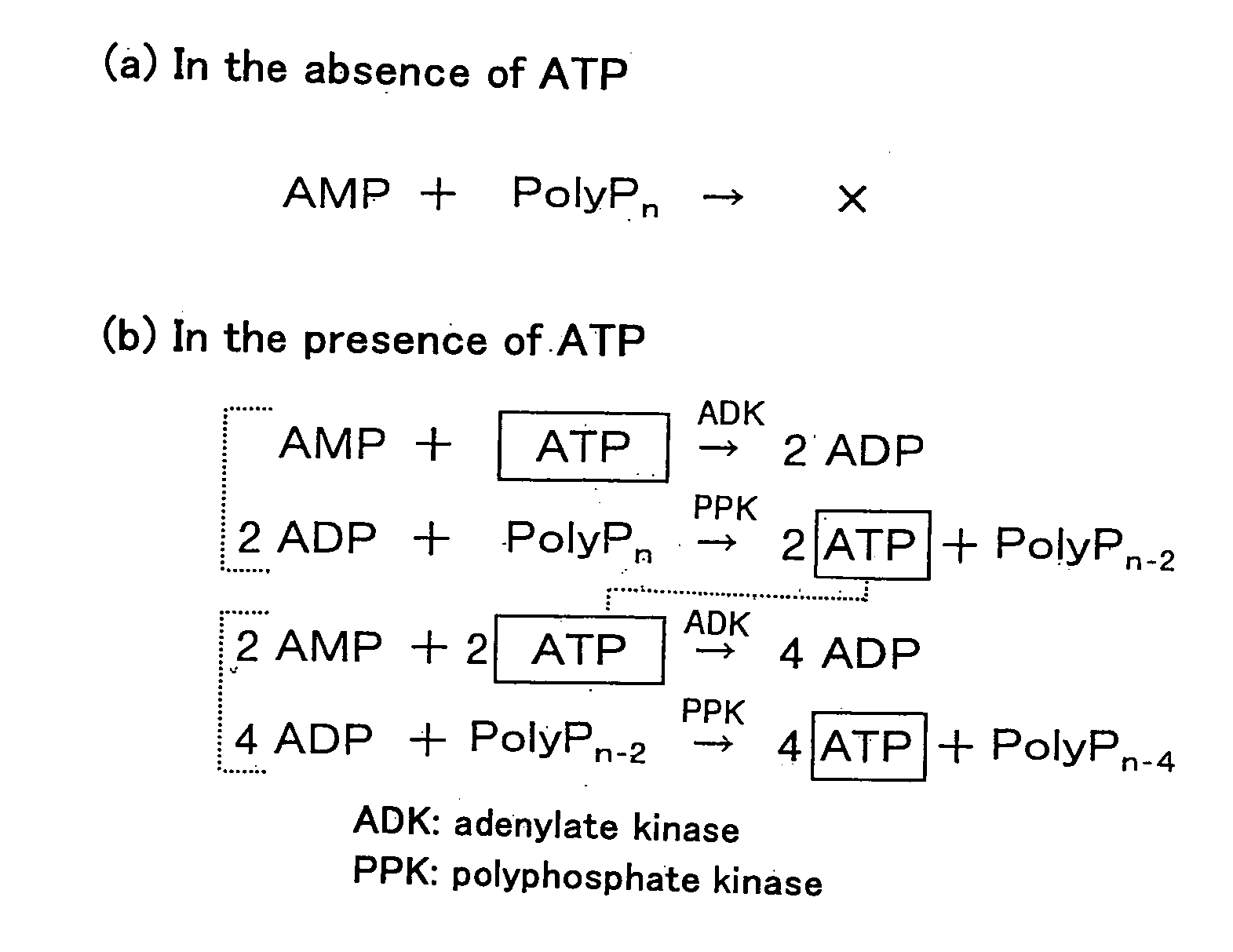
Method of Amplifying Atp and Use Thereof
a technology of amplifying atp and amplifying atp, which is applied in the field of amplifying atp, can solve the problems of insufficient sensitivity for industrial or practical applications, detection limits of conventional methods for assaying atp, and inability to use methods in practice, etc., and achieves the effect of rapid detection
- Summary
- Abstract
- Description
- Claims
- Application Information
AI Technical Summary
Benefits of technology
Problems solved by technology
Method used
Image

Examples
example 1
Preparation of PPK-ADK
[0047]Primers for obtaining a gene (ppk) encoding E. coli polyphosphate kinase (see Akiyama, M. et al., “The polyphosphate kinase gene of Escherichia coli. Isolation and sequence of the ppk gene and membrane location of the protein,” J. Biol. Chem., vol. 267, pp. 22556-22561 (1992)) are as follows:
(SEQ ID No: 1)GGATCTAGATGAATAAAACGGAGTAAAAGTand(SEQ ID No: 2)GGAGGATCCGCCGCCGCCGCCTTCAGGTTGTTCGAGTGATTT.
[0048]The primer of SEQ ID No: 1 has a sequence for introducing a restriction enzyme XbaI recognition site in the 5′ terminal of the ppk gene. SEQ ID No: 2 is designed so that four glycines are attached to the C-terminal of the PPK, and further has a sequence for introducing a restriction enzyme BamHI recognition site in the 3′ terminal.
[0049]Primers for obtaining a gene (adk) encoding E. coli adenylate kinase gene (Brune, M. et al., “Cloning and sequencing of the adenylate kinase gene (adk) of Escherichia coli,” Nucleic Acids Res., vol. 13, pp. 7139-7151 (1985)) ar...
example 2
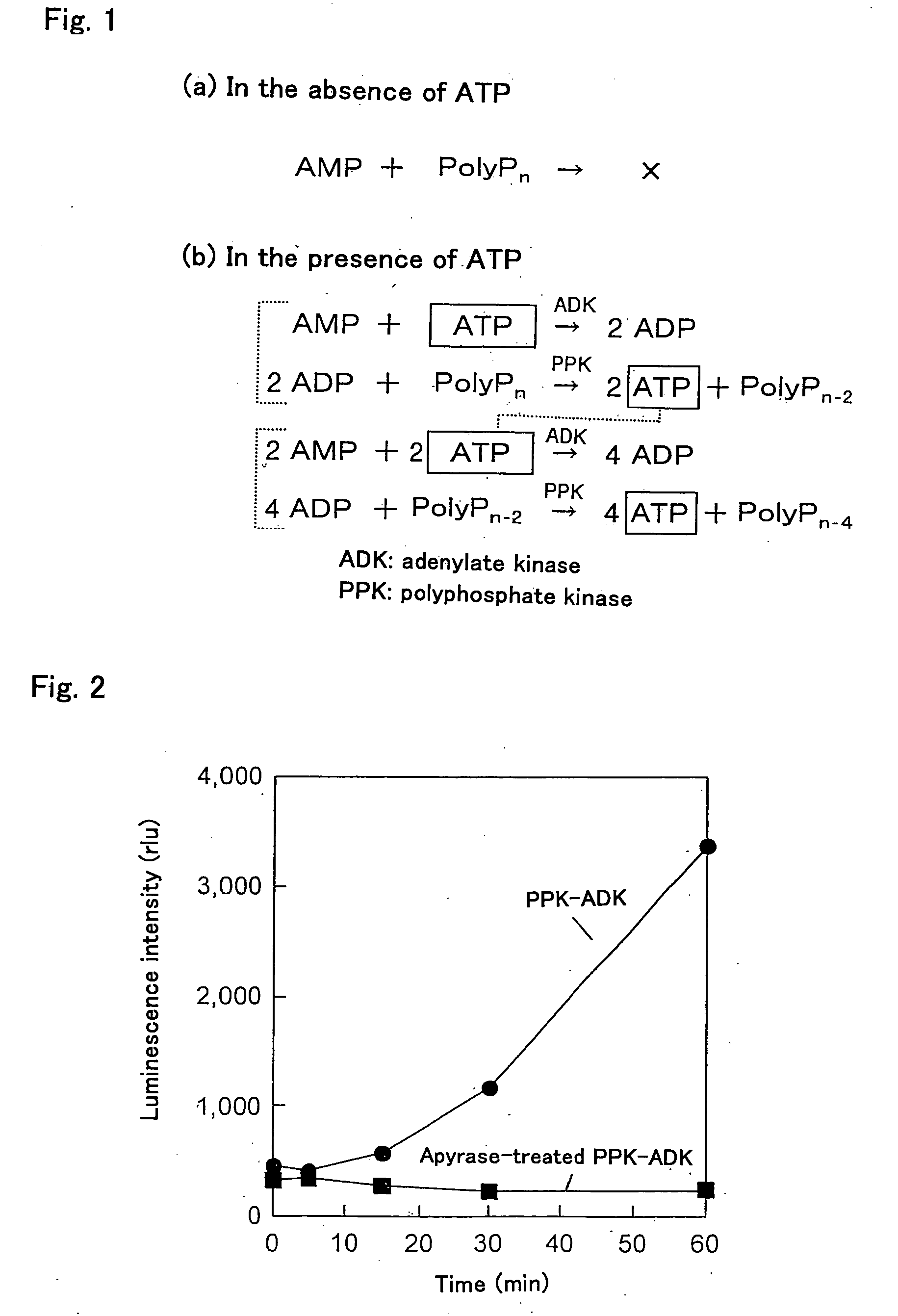
Removal of ADP Bound to PPK-ADK
[0057]In order to remove the ADP, which was an impurity bound to the PPK-ADK obtained in Example 1, 180 μg of the PPK-ADK were reacted with apyrase (200 U) for one hour in the presence of 60 mM Tris-HCl (pH 8), 8 mM MgCl2 and 10 mM polyphosphate. After the reaction was finished, the PPK-ADK from which the ADP was removed was collected by using a Hitrap chelating column again. Hereinafter, this PPK-ADK is referred to as the “apyrase-treated PPK-ADK”. It should be noted that one unit of apyrase releases 1 μmol of phosphate from ATP or ADP per minute at 30° C.
[0058]Next, 50 μl of a reaction mixture containing 0.16 μg of the apyrase-treated PPK-ADK, 10 μM AMP, 400 μM polyphosphate, 8 mM MgCl2, and 60 mM Tris-HCl (pH 7.4) were prepared. Then, 5 μl of the reaction mixture were sampled and mixed with 40 μl of an ATP bioluminescence assay reagent (Roche), and luminescence was measured immediately by using a multiplate luminometer (ARVO, Wallac).
[0059]As shown ...
example 3
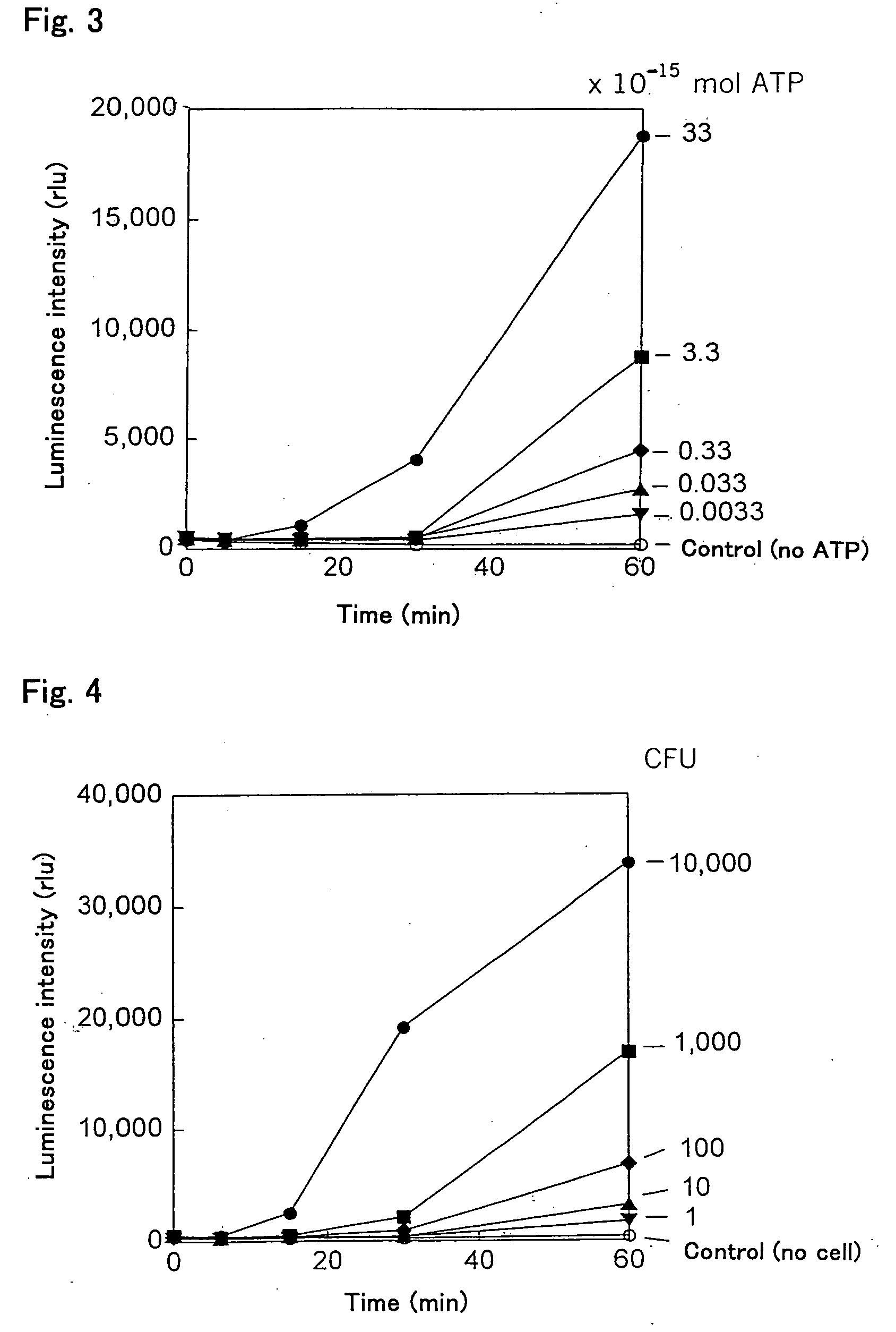
Ultrasensitive Bioluminescence Assay
[0061]First, 48 μl of a reaction mixture containing 0.16 μg of the apyrase-treated PPK-ADK, 10 μM AMP, 400 μM polyphosphate, 8 mM MgCl2, and 60 mM Tris-HCl (pH 7.4) were prepared, and then 2 μl of an ATP sample were added to this reaction mixture to amplify ATP. Thereafter, 5 μl of the reaction mixture were sampled over time and mixed with 40 μl of an ATP bioluminescence assay reagent, and luminescence was measured immediately by using a multiplate luminometer. For comparison, a sample was prepared without amplifying ATP (without adding the PPK-ADK), and the luminescence thereof was measured. Each value of luminescence is the mean ±standard deviation of three different measurements. The increase in luminescence over time is shown in FIG. 3, and the results of ATP amplification after 60 minutes are shown in Table 1.
TABLE 1Luminescence (rlu)ATP amplificationATP (fmol)WithoutWith330813 ± 2228,180 ± 160633113 ± 1418,793 ± 241 3.350 ± 68,767 ± 4430.335...
PUM
 Abstract
Description
Claims
Application Information
 - R&D
- Intellectual Property
- Life Sciences
- Materials
- Tech Scout
- Unparalleled Data Quality
- Higher Quality Content
- 60% Fewer Hallucinations
Browse by: Latest US Patents, China's latest patents, Technical Efficacy Thesaurus, Application Domain, Technology Topic, Popular Technical Reports.
© 2025 PatSnap. All rights reserved.Legal|Privacy policy|Modern Slavery Act Transparency Statement|Sitemap|About US| Contact US: help@patsnap.com