
Nanoparticles for Imaging Atherosclerotic Plaque

a technology of nanoparticles and plaques, applied in the field of coating nanoparticles, can solve the problems of endothelial dysfunction and modification of lipids, ischemia of distal tissues, myocardial infarction or stroke, etc., and achieve the effects of low dose of contrast agents, low dose, and high specificity

Inactive Publication Date: 2008-08-28
RGT UNIV OF CALIFORNIA
View PDF4 Cites 16 Cited by
  • Summary
  • Abstract
  • Description
  • Claims
  • Application Information

AI Technical Summary

Benefits of technology

[0009]The contrast agent of the present invention is coupled to ligands that are recognized by macrophage specific receptors to develop a targeted contrast agent. Since the migration of macrophages into a disease tissue is a dynamic process, utilization of receptors on immune cells enables contrast imaging of the progression of the disease and because of the specificity, enables low doses of contrast agent to be used. The ability to track the progression of the disease with high specificity and low dose (of contrast agent) could lead to a greater understanding of disease progression and aid in development of therapeutics.

Problems solved by technology

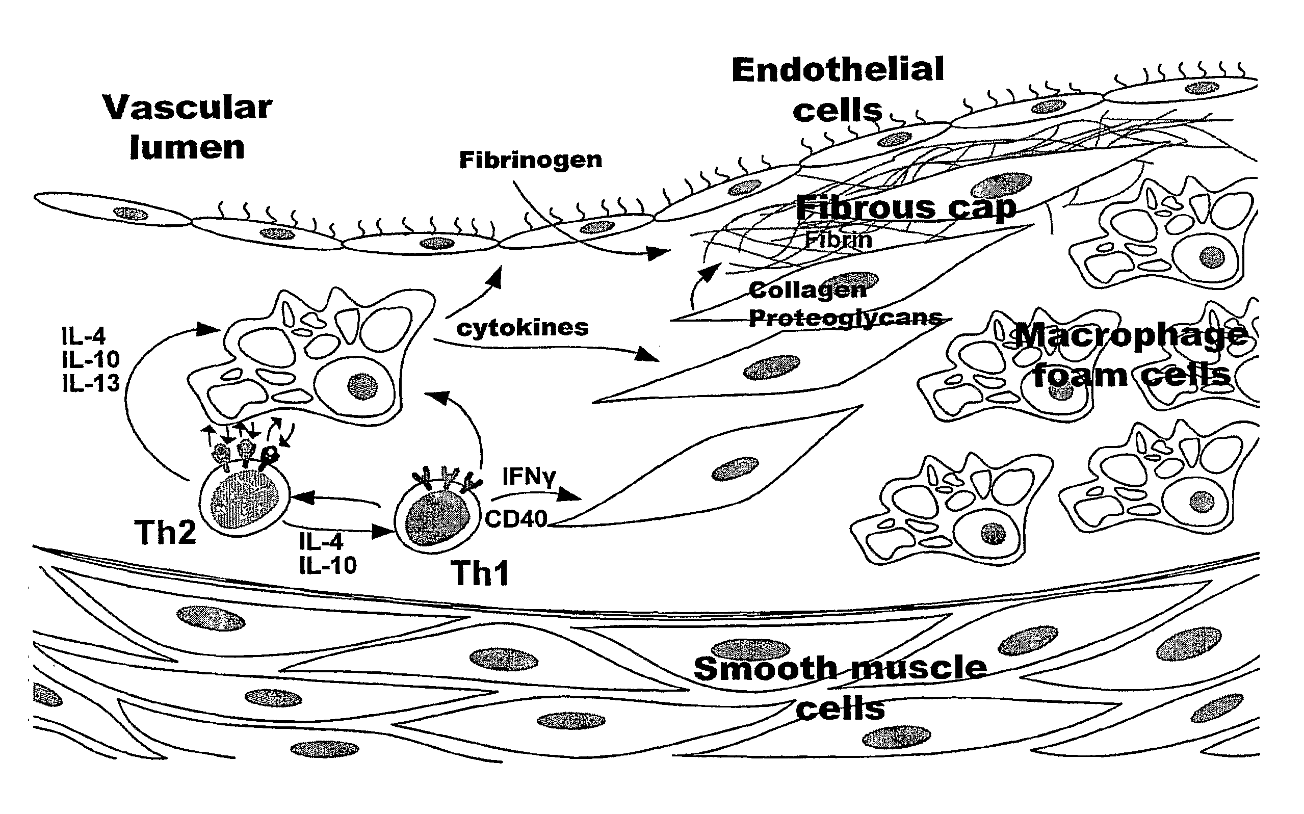
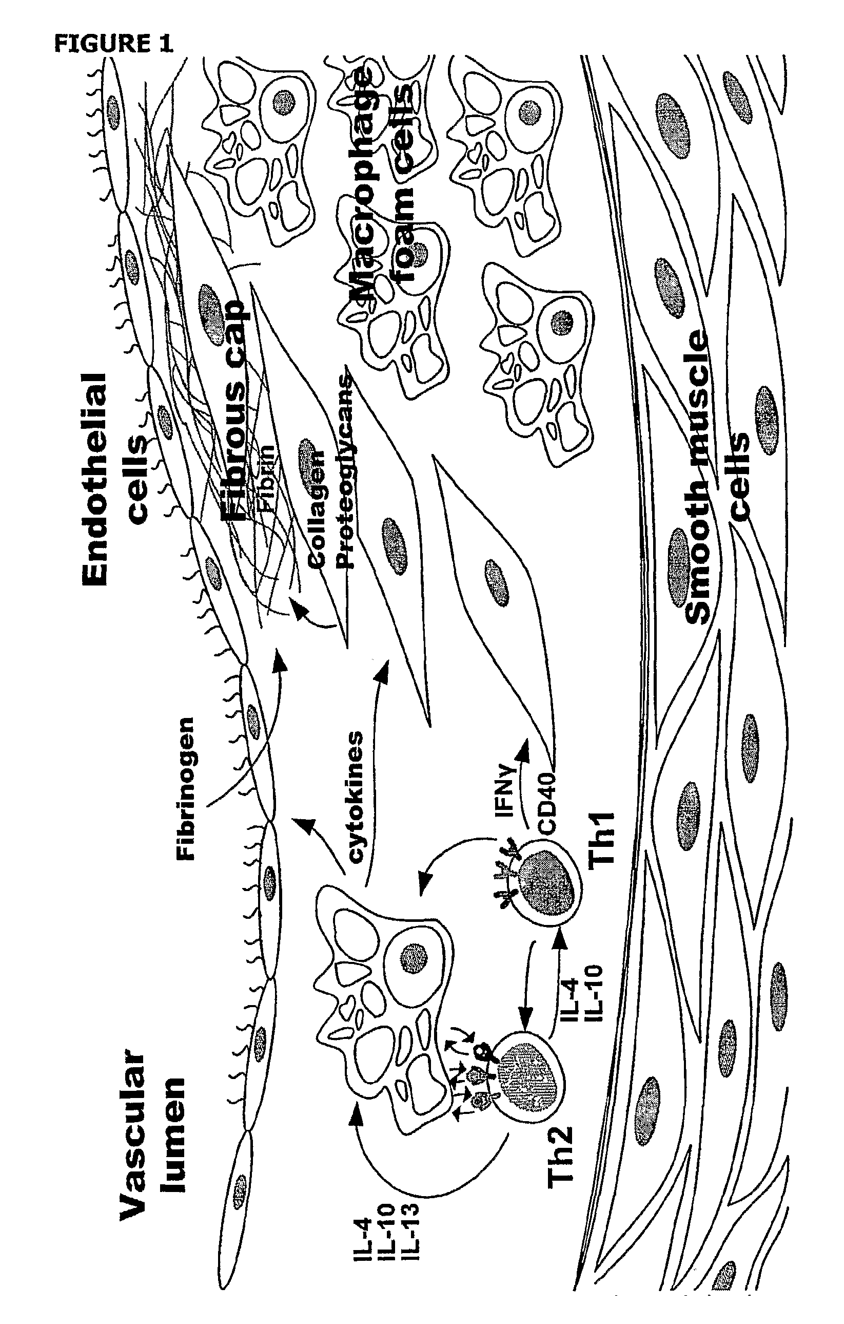
Oxidative stress due to poor diet, smoking, irregular flow at bifurcations and stress can lead to endothelial dysfunction and modification of lipids, specifically low-density lipids (LDL).
The plaque can then extend into the lumen and obstruct blood flow, eventually leading to ischemia of distal tissues.
Or the fibrous cap can become weakened due to immune cell activity and rupture, forming embolisms that can occlude smaller vessels of the heart or brain, leading to myocardial infarction or stroke, respectively.
The current gold standard for detecting atherosclerosis, angiography, is only capable of detecting stenosis, which yields no information about plaque development within the vessel wall.
However, these targeted agents are for markers that are expressed at advanced stages of the disease, not the initial development.
Current imaging techniques are not sophisticated enough for the detection of early plaque components and early plaque development.

Method used


Image

  • Nanoparticles for Imaging Atherosclerotic Plaque
  • Nanoparticles for Imaging Atherosclerotic Plaque
  • Nanoparticles for Imaging Atherosclerotic Plaque

Examples


example 1

Dextran Sulfate Coated Iron Oxide Nanoparticles Created by Layering

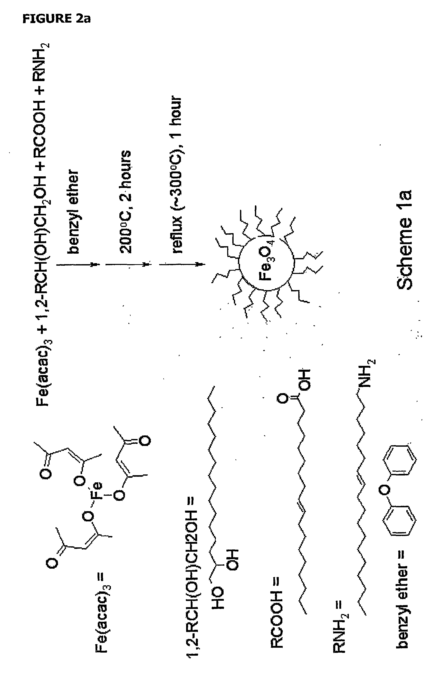
[0061]The iron oxide core was synthesized using a method by Sun and colleagues (Sun et al., 2004) for synthesis of oleic acid / oleylamine coated particles that allows for precise control of particle size. Control of particle size is useful for modeling relaxation properties of the particles and tailoring optimal contrast agent design, as relaxation is size dependent (Yung et al., 2003, Koenig et al., 2002, Roch et al., 1999, Koenig et al., 1995). The general synthesis is shown in scheme 1 (FIG. 2), in which the iron oxide core is formed, transferred to water, and then coated with dextran sulfate via a layer-by-layer (LbL) technique. Magnetite cores were formed using a protocol by Sun (Sun et al., 2004) in which an iron precursor is oxidized to form 6 nm iron oxide. The oleic acid / oleylamine stabilized iron oxide particles were then transferred to water using tetramethylammonium hydroxide (TMAOH) (Euliss et al., 2003)....

example 2

Dextran Sulfate Doped Iron Oxide Nanoparticles

[0063]The initial dextran coated particle synthesis (Palmacci et al., 1993, Paul et al., 2004) was altered to include a small amount of dextran sulfate mixed with reduced dextran (rd) to form DS-doped-rdUSPIOs. Smaller particles less than 50 nm may be ideal as their smaller size will increase circulation time and reduced clearance by the reticuloendothial system (Pratten et al., 1986, Bowen et al, 2002).

[0064]We modified the USPIO synthesis proposed by Paul and colleagues (Paul et al., 2004) to include a small proportion (˜5%) of dextran sulfate mixed in with the reduced dextran. The general core synthesis is as follows: FeCl2+2FeCl3→[Fe(OH)2+2Fe2O3.dextran (or dextran sulfate)]→Fe3O4.dextran / dextran sulfate; where the brackets represent an intermediate step (Thomassen et al., 1991). Using a very small percentage of dextran sulfate may allow the iron oxide cores to form with a small amount of sulfate groups attached for recognition by th...

example 3

Silica Coated Iron Oxide Nanoparticles

[0065]Synthesis

[0066]Silica coated particles were synthesized. Silica particles have been widely used for stabile nanoparticles platforms as they are stabile in a wide pH range (Klotz et al., 1999) and silica, due to its polyanionic nature, has been shown to be recognized by the macrophage SR (Platt et al., 2001). We show here that silica coated particles are recognized by macrophages and can be used to label atherosclerotic plaques. The general synthetic route is shown in scheme 2 (FIG. 4). The iron oxide cores were again synthesized according to the Sun protocol (Sun et al, 2004), and transferred to water as before (Euliss et al., 2003). Silica coated particles were then made by the absorption of Si onto the iron oxide by base hydrolysis of tetraethylorthosilicate (TEOS) (Lu et al., 2002).

[0067]FIG. 5 is a TEM image of 80 nm Silica coated particles demonstrating a 10 nm iron oxide core. Dynamic light scattering showed an overall particle diame...


PUM

PropertyMeasurementUnit
particle sizeaaaaaaaaaa

Abstract

Atherosclerosis is an inflammatory disease of the arterial walls and represents a significant health problem in developed nations. Described is a targeted Magnetic Resonance Imaging (MRI) contrast agent for in vivo imaging of early stage atherosclerosis. Early plaque development is characterized by the influx of macrophages, which express a class of surface receptors known collectively as the scavenger receptors (SR). The macrophage scavenger receptor class A (SRA) is highly expressed during early atherosclerosis. The macrophage SRA therefore presents itself as an ideal target for labeling of lesion formation. By coupling a known ligand for the scavenger receptor, dextran sulfate, to a MRI contrast agent, early plaque formation can be detected in vivo. Targeted MR contrast agents offer a unique opportunity to visualize early plaque development in vivo with high sensitivity and resolution, allowing or early diagnosis and treatment of atherosclerosis.

Description

CROSS REFERENCE TO RELATED APPLICATIONS[0001]This application claims the benefit of U.S. Provisional Application 60 / 582,768, filed Jun. 25, 2004 which is hereby incorporated by reference in its entirety.FIELD OF THE INVENTION[0002]The compositions and methods described herein generally relate to coated nanoparticles used for the detection of macrophages and inflammatory diseases such as atherosclerosis.BACKGROUND OF THE INVENTION[0003]Heart disease is one of the leading killers in developed nations. In the United States alone there are approximately 5 million Americans living with heart disease with 550,000 new cases each year. Furthermore, roughly three quarters of the million cardiovascular disease (CVD) deaths each year are due to atherosclerosis (Heart Disease and Stroke Statistics—2005 Update, American Heart Association: American Heart Association; 2005. 1-63 p.), an inflammatory disease of the arterial vessel wall. Early diagnosis of atherosclerosis would allow for early treat...

Claims


Application Information

Patent Timeline
Patent Type & Authority Applications(United States)
IPC IPC(8): A61K49/18A61K49/00A61P9/10A61K49/06
CPCA61K49/183B82Y5/00A61K49/1863A61P9/10
Inventor LOUIE, ANGELIQUE Y.JARRETT, BENJAMIN R.
Owner RGT UNIV OF CALIFORNIA
Learn More