Eureka AIR delivers breakthrough ideas for toughest innovation challenges, trusted by R&D personnel around the world.

Endoscope Propulsion System and Method

a propulsion system and endoscope technology, applied in the field of endoscopy, can solve the problems of too insensitive test for effective screening, early colon cancer and polyps are asymptotic, and about 10 tests too insensitive to effective screening, so as to improve the safety of colonoscopy, reduce procedure-related pain, and safe and effective colon cancer screening method

Inactive Publication Date: 2008-07-31
BERN M JONATHAN +1
View PDF10 Cites 91 Cited by
  • Summary
  • Abstract
  • Description
  • Claims
  • Application Information

AI Technical Summary

Benefits of technology

[0020]One advantage provided by the present invention is a safe and effective low cost method for colon cancer screening. To achieve this end, the invention provides an endoscopic propulsion unit that can attach to currently available colonoscopes. The endoscopic propulsion unit can advance a colonoscope in the colon lumen without stretching the colon wall, greatly reducing procedure-related pain. An additional advantage provided by the invention relates to safety. For example, safety of colonoscopy is improved through the use of the present invention by reducing or eliminating the risk of colon perforation. In contrast to other propulsion units, the endoscopic propulsion unit of the present invention advances a colonoscope by pulling the distal end of the instrument. This reduces the likelihood of perforations, and reduces the amount of pain experienced by the patient. Furthermore, the present invention allows relatively painless colonoscopy that can be performed safely in a physician's office. By removing the need for high level sedation, colonoscopy can now be moved to a lower cost center, such as a physician's office or outpatient clinic. This movement away from hospital settings could result in a 66% or greater savings in the total colonoscopy cost. This comfortable, effective, affordable and safe method for colon cancer screening provided by the present invention can be widely used to reduce colon cancer mortality. Other advantages will be realized through consideration of the following disclosure and practice of the invention.
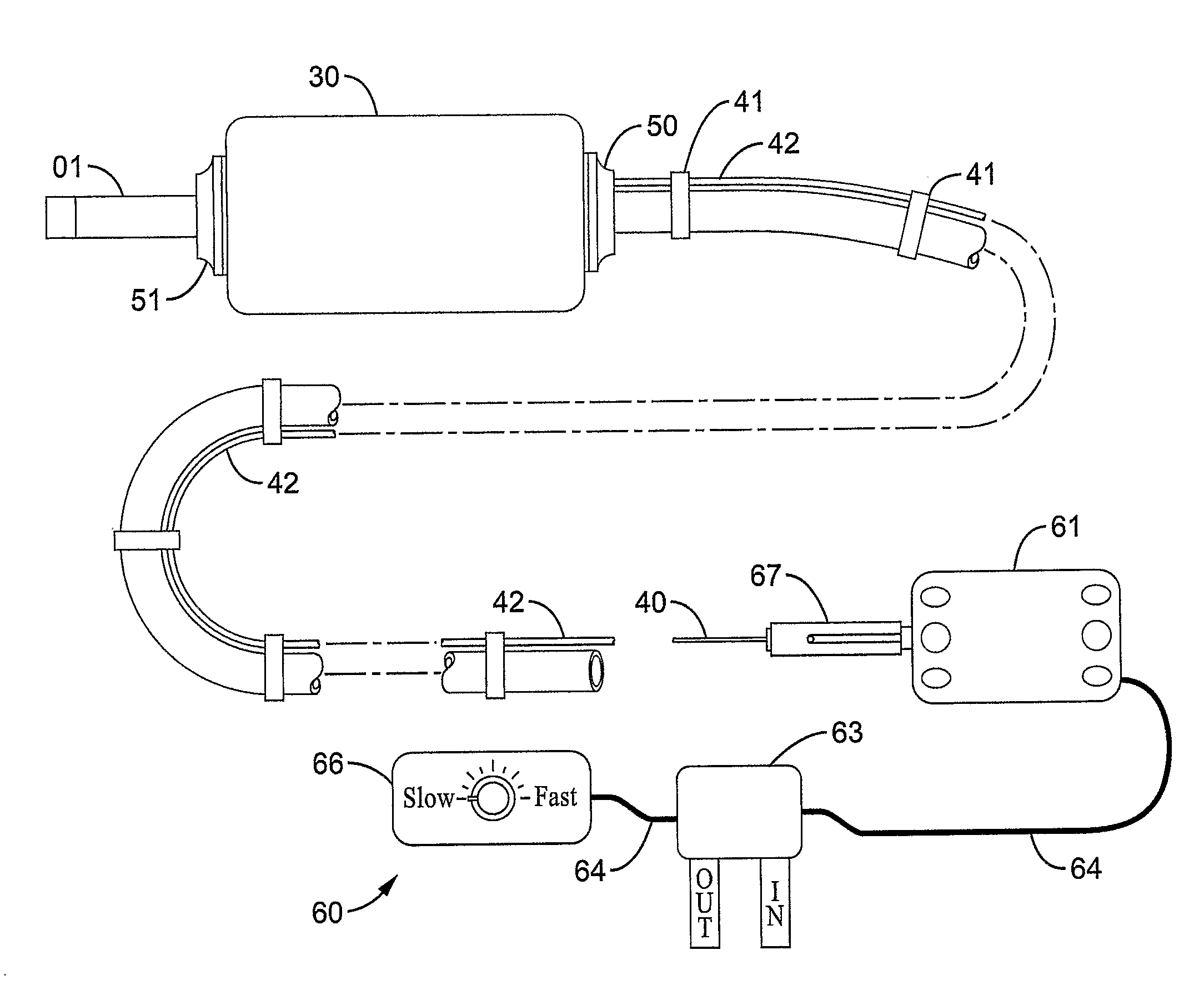
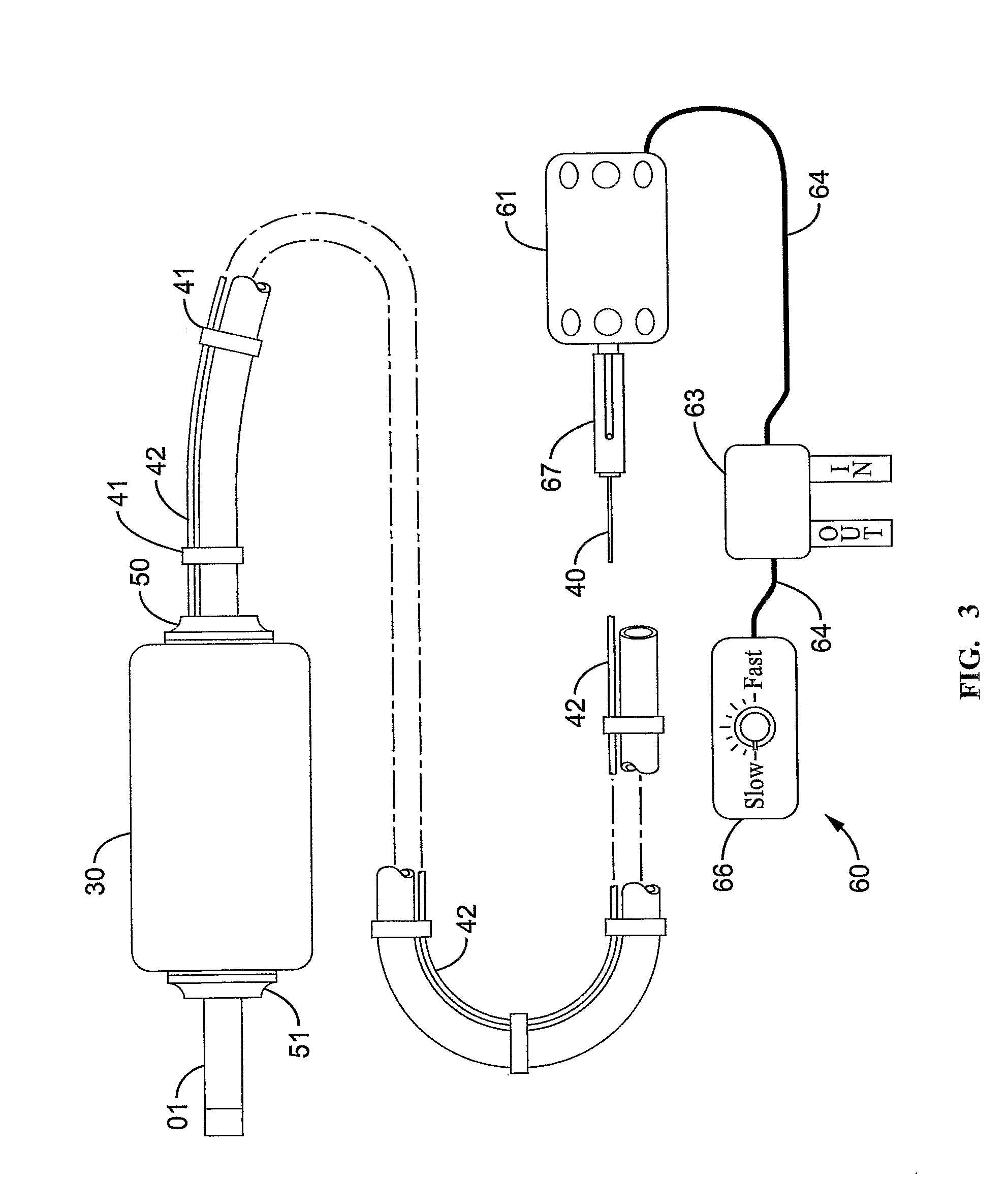
[0021]In a first aspect, the invention provides a device, such as one for use with a medical instrument. The device is capable of self-propelled motion through cavities defined by one or more walls, such as pipes and tubes, and such as body spaces, cavities, lumens, etc. (used interchangeably herein to denote an area within an animal, including human, body that is defined and bordered by a wall). When attached to another instrument, such as a medical instrument, provides the instrument with the ability to move through the cavities, such as body spaces, substantially without propulsive force provided by a human, or with relatively little human force. In general, the device comprises a drive unit or transmission for converting rotational energy from a drive shaft into longitudinal (i.e., forward or backward) movement of the device along a cavity. The drive unit comprises means for receiving one or more drive shafts, such as a drive shaft receptacle; means for converting rotational force provided by the drive shaft to longitudinal force, such as a radial gear, a series of interconnecting gears, or a worm gear; means for providing the longitudinal force of the drive unit to an exterior surface of the drive unit to cause the drive unit to move longitudinally, such as a rotatable rod or band comprising a suitable surface; and means for translating the longitudinal force of the drive unit to longitudinal force exerted against a cavity surface to cause the drive unit to move longitudinally along the cavity, such as a membranous element comprising a surface that releasably contacts the means for providing longitudinal force to a surface of the drive unit and releasably contacts the cavity surface. As can be seen, the device of the invention comprises two sub-parts that can be provided separately but combined to function together. That is, the drive unit may be provided with or without the means for translating longitudinal force from the drive unit to the cavity surface; where the two are provided separately, they can be combined to provide a unitary device.

Problems solved by technology

Unfortunately early colon cancer and polyps are asymptotic.
In part because of the limitations of these tests, only about 10% of the United States population is currently screened for this common preventable cause of death.
Unfortunately only about 30% of colon cancers can be detected by fecal occult blood testing, making this test too insensitive for effective screening.
Unfortunately flexible sigmoidoscopy is too uncomfortable for many patients to tolerate.
Flexible sigmoidoscopy is painful because the scope is advanced in the colon by pushing the semi-rigid tube against the colon wall.
Stretching of the colon causes intense visceral pain.
In addition to pain, stretching the colon too far can result in colon perforation, a potentially life threatening complication of flexible sigmoidoscopy.
Unfortunately colonoscopy is far too uncomfortable to be performed without high level intravenous sedation or general anesthesia.
With the added operating room charges colonoscopy becomes quite costly.
Several such disclosures involve robotic endoscopes that are generally complex devices with multiple interacting segments.
Furthermore, the examining physician can not directly assist in the movement of the scope in an organ lumen.
The lack of direct physician control will markedly increase the risks of robotic endoscopy.
Even the slightest malfunction of the complex control mechanism could cause devastating complications for a patient.
Thus high inflation pressures may be required to grip and fix the toroidal balloon to the organ wall.
Therefore, robotic endoscopy according to these prior devices and methods may often require high level sedation or general anesthesia to permit a comfortable examination.
In this regard, robotic endoscopy according to these prior disclosures offers no additional benefits to currently available endoscopic procedures.
Furthermore, the extensor module of these prior robotic endoscope disclosures is constantly changing the axial length of the robotic endoscope.
As the robotic endoscope is constantly changing length, currently available endosurgical devices, such as biopsy forceps or polypectomy snares, may be very difficult if not prevented from conjunctive use.
The mechanical complexity of this prior approach and the need for computer control systems generally relate to relatively high production cost for the robotic endoscope.
And, as in many fields, high production cost could substantially limit the availability of robotic endoscopy for widespread clinical use, such as in colorectal cancer screening.
Moreover, sufficiently high production cost might also prohibit disposal of the robotic endoscope after each use.
As disposal would not be generally practical according to these prior approaches, sterilization of the robotic endoscope becomes a likely necessity.
Furthermore, sterilizing such a complex device with multiple mechanical and electronic components would be still a further challenge of substantial difficulty.
The difficulty in sterilizing these robotic endoscopes could result in elevated potential for infectious disease transmission.

Method used


Image

  • Endoscope Propulsion System and Method
  • Endoscope Propulsion System and Method
  • Endoscope Propulsion System and Method

Examples


Embodiment Construction

[0060]Reference will now be made in detail to various exemplary embodiments of the invention, examples of which are illustrated in the accompanying drawings. The following detailed description is provided to detail various elements, combinations, and embodiments of the invention, and is not intended as a limitation of the invention to the particular elements, combinations, and embodiments exemplified.

[0061]In a first aspect, the invention provides a device, such as a medical device or a device for use in non-medical situations. The device may be used for diagnostic purposes and therapeutic purposes in its medical embodiments, and for diagnostic and reparative purposes in its non-medical embodiments. In its various embodiments, it is appropriately sized to fit and function within the particular cavity it is to be used in. Thus, in embodiments, it is sized to fit in a human cavity, such as a colon, vein, or the like. It also may be used in conjunction with another instrument of device...


PUM


Abstract

The present invention provides a system and method for active propulsion of devices, such as endoscopes, along cavities, such as body lumens. The propulsion system can be attached to a commercially available endoscope, or be provide affixed together, and moves the endoscope in a lumen by pulling it forward. The present invention further provides a method of diagnosing diseases and disorders, and treatment of diseases and disorders, using a device according to the invention.

Description

CROSS-REFERENCE TO RELATED APPLICATIONS[0001]The present application relies on, and claims the benefit of the filing date of, U.S. patent application Ser. No. 11 / 140,595, filed 27 May 2005. Priority of the filing date of this application is claimed, and the disclosure of the application is incorporated herein by reference in its entirety.BACKGROUND OF THE INVENTION[0002]1. Field of the Invention[0003]The present invention relates to the field of health care. More specifically, the invention relates to the field of endoscopy, and particularly to devices and methods for performing endoscopic examinations and surgeries.[0004]2. Description of Related Art[0005]Each year, 60,000 Americans die from colon cancer, making colon cancer the second leading cause of cancer death in the United States. Early detection of the disease greatly improves survival. Furthermore, removal of pre-cancerous polyps can be achieved endoscopically, which prevents colon cancer altogether. Unfortunately early col...

Claims


Application Information

Patent Timeline
Patent Type & Authority Applications(United States)
IPC IPC(8): A61B1/00
CPCA61B1/0016A61B1/00148
Inventor BERN, M. JONATHANPEACOCK III, JAMES C.
Owner BERN M JONATHAN
Why Eureka
Eureka Blog
Learn More