Non-invasive methods and related compositions for identifying compounds that modify in vivo aggregations of disease-related polypeptides

Inactive Publication Date: 2008-07-10
UNIV OF WASHINGTON
View PDF0 Cites 1 Cited by
  • Summary
  • Abstract
  • Description
  • Claims
  • Application Information

AI Technical Summary

Benefits of technology

[0008]Embodiments of the present invention are directed to various high-throughput methods for identifying active compounds that are likely to permeate the blood-brain barrier (“BBB”), and that are useful for the treatment of various neurodegenerative diseases, including the Huntington's Disease (HD), Parkinson's Disease (PD), Alzheimer's Disease (AD), Amyotrophic Lateral Sclerosis (ALS), and related diseases. The pathogenesis of many neurodegenerative diseases leads to the aggregation of endogenous proteins, including self-aggregating polypeptide variants, that are expressed within compartments of central nervous tissues, including brain neurons. In various embodiments, methods and compositions of the present invention enable the exogenous expression of one or more neurodegenerative-disease-related polypeptide variants within the ocular lens of an animal host. The formation of aggregates containing neurodegenerative-disease-related polypeptide variants increases the opacity of the lens, in a manner similar to the development of age-onset cataracts. The effect of a test compound in decreasing aggregate formation and / or in destabilizing aggregates that contain neurodegenerative-disease-related polypeptide variants can be visually monitored and quantified in living animal hosts by employing conventional cataract-detecting instrumentation and related methods. Compounds identified by assays of the present invention that can permeate the blood-ocular barrier can be predictive of the likelihood that the identified compounds are also permeable across the BBB. Animal hosts that can express various neurodegenerative-disease-related polypeptides in both the ocular lens and the central nervous tissues are provided.

Problems solved by technology

However, determining the bioavailability of drugs into the brain is experimentally challenging.
Furthermore, most drugs do not pass the BBB due to improper size, charge, and / or other structural properties, as described below.
The characterization for the bioavailability of each active compound is very time-consuming, and the evaluation of a large number of compounds utilizing such methods is impractical.
Histological examinations of brain sections removed from diseased-animal hosts are not only invasive, but such examinations are very labor-intensive and impractical for the evaluation of numerous compounds.

Method used


Image

  • Non-invasive methods and related compositions for identifying compounds that modify in vivo aggregations of disease-related polypeptides
  • Non-invasive methods and related compositions for identifying compounds that modify in vivo aggregations of disease-related polypeptides
  • Non-invasive methods and related compositions for identifying compounds that modify in vivo aggregations of disease-related polypeptides

Examples


example 1

Construction of Transgenic Mice Expressing Huntingtin-Exon-1 Variants within the Ocular Lens

[0120]Transgenic mice that can express mutant huntingtin fragments in the ocular lens can be employed for screening compounds to identify active compounds for the prevention, management, and / or treatment of Huntington's disease and related diseases. FIG. 8 is a schematic of an exemplary expression cassette for expressing a gene of interest within the ocular lens of an animal host. In FIG. 8, various forms of EGFP-tagged, huntingtin-exon-1 fragments containing variable lengths of poly-Q-repeat domains can be produced from an expression construct 800 that includes a crystallin promoter 802 positioned upstream and operably-linked to a huntingtin fragment containing an expanded-CAG repeat, such as “Htt Exon1+Qn”804, which is positioned upstream and operably-linked to an EGFP-encoding sequence 808. Optionally, a 3′ untranslated sequence, such as a “SV40-polyA” sequence 810 or an equivalent sequenc...

example 2

Characterization of the Expression of EGFP-Huntingtin-Exon-1 Fragments within the Ocular Lens of Animal Hosts by Slit-Lamp Biomicroscopy

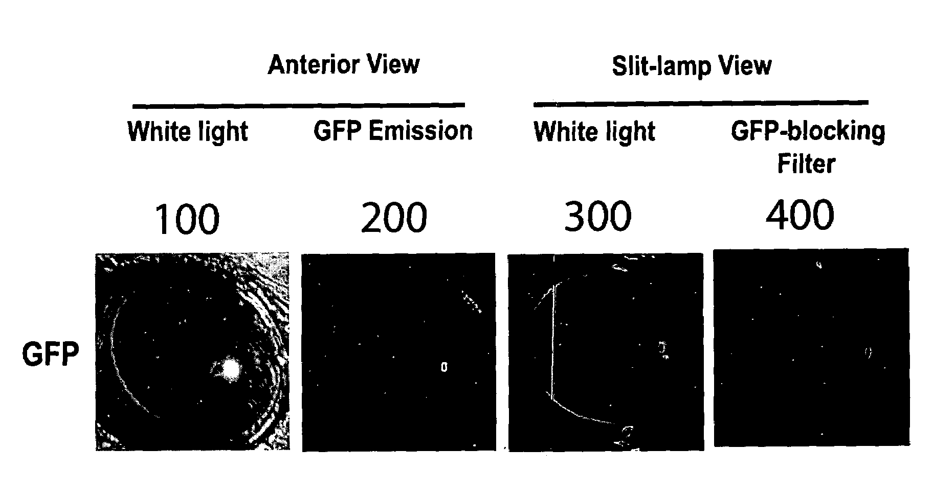
[0124]The transgenic animals described in Example 1 are evaluated to quantify poly-Q-mediated-aggregation within the ocular lens of an animal host. In three of the four founder lines, robust lens-specific, or lenticular, expression is observed by employing a slit-lamp biomicroscope that permits the viewing of an optical section of the lens and the digital imaging of GFP fluorescence that arise from the lens of each animal host. FIG. 10 is a digital image of the lens of transgenic founder mice expressing huntingtin-exon-1 variants that contain extended poly-glutamine (Q) domains. FIG. 10A is a digital image of an anterior view of a hypothetical animal host illuminated with white light. FIG. 10B is a digital image of an anterior view of a hypothetical animal host that records the filtered light emitted by GFP-tagged proteins within the ocular lens, un...

example 3

Microscopic Analysis of Inclusion Bodies Containing Mutant Huntingtin Fragments

[0126]FIG. 11A is a microscopic view of lens that expresses a mutant huntingtin fragment containing a non-extended poly-Q domain “25Q,” removed from a transgenic mouse at 8 weeks of age at 10× magnification. In FIGS. 11A-11C, and 11F, GFP florescence appears in green, and the nuclei stained with propidium iodide stains in red. FIG. 11B illustrates an exemplary lens section removed from a transgenic mouse that shows a thin halo (green) of inclusion bodies at 3 weeks. FIG. 11C illustrates an exemplary lens section removed from a transgenic mouse expressing a mutant huntingtin “72Q” fragment at 8 weeks that shows a ten-fold increase in the number of inclusion bodies within the halo region (green) during this time period. FIG. 11D illustrates an exemplary lens section of lens removed from a transgenic mouse expressing a mutant huntingtin “72Q” fragment examined for GFP fluorescence at 20× magnification. In FI...


PUM

PropertyMeasurementUnit
Fractionaaaaaaaaaa
Fluorescenceaaaaaaaaaa
Bioavailabilityaaaaaaaaaa

Abstract

Embodiments of the present invention are directed to various high-throughput methods for identifying compounds that are useful for the treatment of various neurodegenerative diseases, including the Huntington's Disease (HD), Parkinson's Disease (PD), Alzheimer's Disease (AD), and Amyotrophic Lateral Sclerosis (ALS). Methods and compositions of the present invention enable the exogenous expression of one or more neurodegenerative-disease-related polypeptide variants within the ocular lens of an animal host. The formation of aggregates containing neurodegenerative-disease-related polypeptide variants increases the opacity of the lens, in a manner similar to the development of age-onset cataracts. The effect of a test compound in decreasing aggregate formation and / or destabilizing aggregates that contain neurodegenerative-disease-related polypeptide variants can be visually monitored and quantified in living animal hosts by employing conventional cataract-detecting instrumentation and related methods.

Description

TECHNICAL FIELD[0001]The present invention relates to compositions and methods for screening compounds in order to identify active compounds that can modify the in vivo formation of aggregates containing various disease-related polypeptides and that are therefore useful for the prevention, management, and / or treatment of various neurodegenerative diseases.BACKGROUND OF THE INVENTION[0002]To protect the central nervous system from noxious agents, many animals, including humans, have developed various structural barriers, such as the “blood-brain barrier” (“BBB”), so that transport of molecules into the central nervous system can be shielded by passive and active mechanisms. The transport of endogenous and exogenous molecules from the circulatory system into the brain parenchyma is regulated mainly by the BBB. The BBB forms a lipophilic membrane consisting of cerebral-capillary-endothelial cells that are tightly arranged so that intercellular junctions between cerebral-capillary-endot...

Claims


Application Information

Patent Timeline
IPC IPC(8): G01N33/00A01K67/00C12N15/63
CPCA01K67/0275A01K2217/05G01N27/447A01K2267/0393C07K14/4747A01K2227/105
Inventor MUCHOWSKI, PAUL J.CLARK, JOHN I.
Owner UNIV OF WASHINGTON
Why Eureka
Try Eureka