
Electromagnetic apparatus for respiratory disease and method for using same

a technology of electromagnetic equipment and respiratory disease, which is applied in the field of electromagnetic equipment for respiratory disease and method for using same, can solve the problems of lasting and deleterious effect on the proper functioning of the apparatus

Inactive Publication Date: 2008-06-05
RIO GRANDE NEUROSCI
View PDF4 Cites 62 Cited by
  • Summary
  • Abstract
  • Description
  • Claims
  • Application Information

AI Technical Summary

Benefits of technology

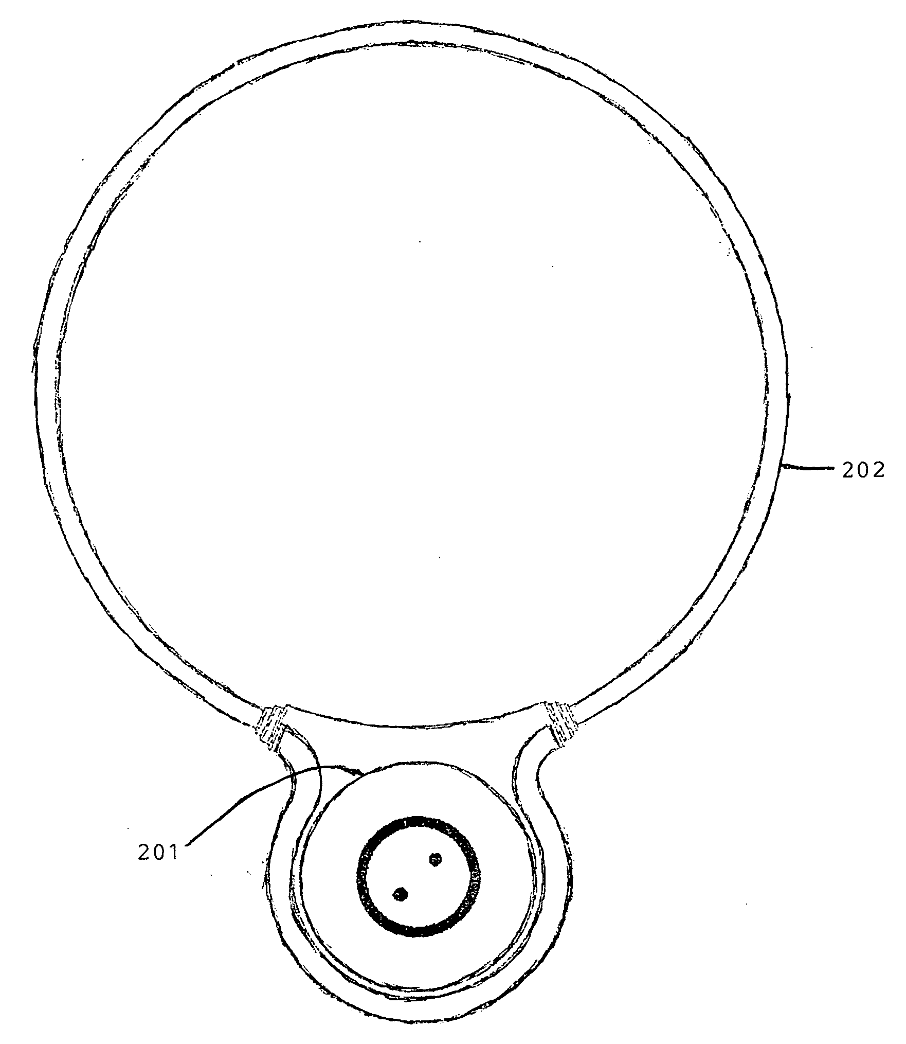
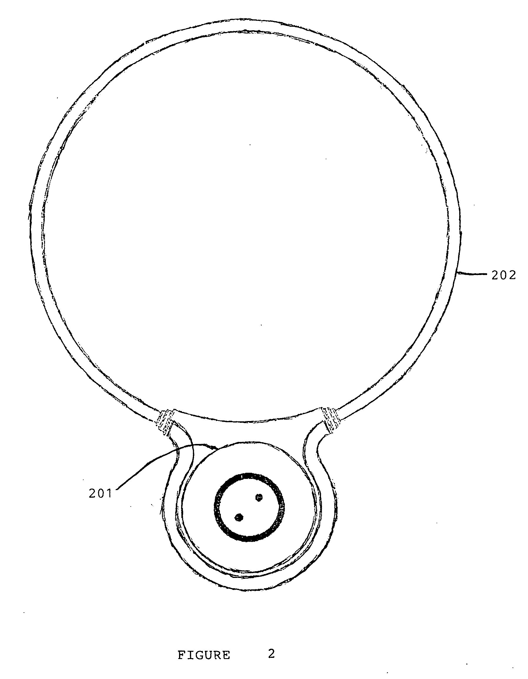
[0087]FIG. 3 depicts a block diagram of an embodiment according to the present invention of a miniature control circuit 300. The miniature control circuit 300 produces waveforms that drive a generating device such as wire coils described above in FIG. 2. The miniature control circuit can be activated by any activation means such as an on / off switch. The miniature control circuit 300 has a power source such as a lithium battery 301. Preferably the power source has an output voltage of 3.3 V but other voltages can be used. In another embodiment according to the present invention the power source can be an external power source such as an electric current outlet such as an AC / DC outlet, coupled to the present invention for example by a plug and wire. A switching power supply 302 controls voltage to a micro-controller 303. Preferably the micro-controller 303 uses an 8 bit 4 MHz micro-controller 303 but other bit MHz combination micro-controllers may be used. The switching power supply 302 also delivers current to storage capacitors 304. Preferably the storage capacitors 304 having a 220 uF output but other outputs can be used. The storage capacitors 304 allow high frequency pulses to be delivered to a coupling device such as inductors (Not Shown). The micro-controller 303 also controls a pulse shaper 305 and a pulse phase timing control 306. The pulse shaper 305 and pulse phase timing control 306 determine pulse shape, burst width, burst envelope shape, and burst repetition rate. In an aspect of the present invention the pulse shaper 305 and phase timing control 306 are configured such that the waveforms configured are detectable above background activity at a target pathway structure by satisfying at least one of a SNR and Power SNR mathematical model. An integral waveform generator, such as a sine wave or arbitrary number generator can also be incorporated to provide specific waveforms. A voltage level conversion sub-circuit 307 controls an induced field delivered to a target pathway structure. A switching Hexfet 308 allows pulses of randomized amplitude to be delivered to output 309 that routes a waveform to at least one coupling device such as an inductor. The micro-controller 303 can also control total exposure time of a single treatment of a target pathway structure such as a molecule, cell, tissue, and organ. The miniature control circuit 300 can be constructed to be programmable and apply a pulsing magnetic field for a prescribed time and to automatically repeat applying the pulsing magnetic field for as many applications as are needed in a given time period, for

Problems solved by technology

In certain respiratory diseases there are physiological deficiencies and disease states that can have a lasting and deleterious effect on the proper functioning of the respiratory system.

Method used


Image

  • Electromagnetic apparatus for respiratory disease and method for using same
  • Electromagnetic apparatus for respiratory disease and method for using same
  • Electromagnetic apparatus for respiratory disease and method for using same

Examples


example 1

[0092]The Power SNR approach for PMF signal configuration has been tested experimentally on calcium dependent myosin phosphorylation in a standard enzyme assay. The cell-free reaction mixture was chosen for phosphorylation rate to be linear in time for several minutes, and for sub-saturation Ca2+ concentration. This opens the biological window for Ca2+ / CaM to be EMF-sensitive. This system is not responsive to PMF at levels utilized in this study if Ca2+ is at saturation levels with respect to CaM, and reaction is not slowed to a minute time range. Experiments were performed using myosin light chain (“MLC”) and myosin light chain kinase (“MLCK”) isolated from turkey gizzard. A reaction mixture consisted of a basic solution containing 40 mM Hepes buffer, pH 7.0; 0.5 mM magnesium acetate; 1 mg / ml bovine serum albumin, 0.1% (w / v) Tween80; and 1 mM EGTA12. Free Ca2+ was varied in the 1-7 μM range. Once Ca2+ buffering was established, freshly prepared 70 nM CaM, 160 nM MLC and 2 nM MLCK w...

example 2

[0096]According to an embodiment of the present invention use of a Power SNR model was further verified in an in vivo wound repair model. A rat wound model has been well characterized both biomechanically and biochemically, and was used in this study. Healthy, young adult male Sprague Dawley rats weighing more than 300 grams were utilized.

[0097]The animals were anesthetized with an intraperitoneal dose of Ketamine 75 mg / kg and Medetomidine 0.5 mg / kg. After adequate anesthesia had been achieved, the dorsum was shaved, prepped with a dilute betadine / alcohol solution, and draped using sterile technique. Using a #10 scalpel, an 8-cm linear incision was performed through the skin down to the fascia on the dorsum of each rat. The wound edges were bluntly dissected to break any remaining dermal fibers, leaving an open wound approximately 4 cm in diameter. Hemostasis was obtained with applied pressure to avoid any damage to the skin edges. The skin edges were then closed with a 4-0 Ethilon ...

example 3

[0102]This example illustrates the effects of PMF stimulation of a T-cell receptor with cell arrest and thus behave as normal T-lymphocytes stimulated by antigens at the T-cell receptor such as anti-CD3.

[0103]In bone healing, results have shown that both 60 Hz and PEMF fields decrease DNA synthesis of Jurkat cells, as is expected since PMF interacts with the T-cell receptor in the absence of a costimulatory signal. This result is consistent with an anti-inflammatory response, as has been observed in clinical applications of PMF stimuli. The PEMF signal is more effective. A dismetry analysis performed according to an embodiment of the present invention demonstrates why both signals are effective and why PEMF signals have a greater effect than 60 Hz signals on Jurkat cells in the most EMF-sensitive growth stage.

[0104]Comparison of dosimetry from the two signals employed involves evaluation of the ratio of the Power spectrum of the thermal noise voltage that is Power SNR, to that of th...


PUM


Abstract

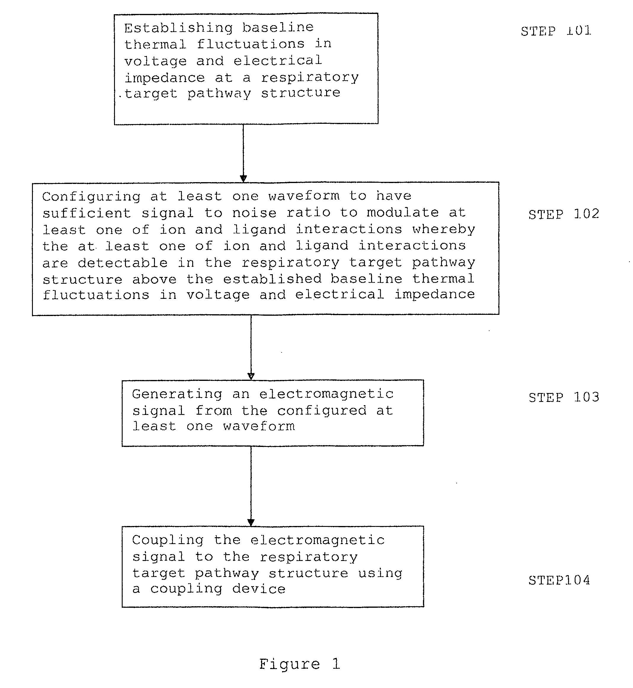
A method for altering the electromagnetic environment of respiratory tissues, cells, and molecules comprising establishing baseline thermal fluctuations in voltage and electrical impedance at a respiratory target pathway structure depending on a state of the respiratory tissue, configuring at least one waveform to have sufficient signal to noise ratio to modulate at least one of ion and ligand interactions whereby the at least one of ion and ligand interactions are detectable in the respiratory target pathway structure above the established baseline thermal fluctuations in voltage and electrical impedance, generating an electromagnetic signal from the configured at least one waveform; and coupling the electromagnetic signal to the respiratory target pathway structure using a coupling device.

Description

[0001]This application claims the benefit of U.S. Provisional Application 60 / 846,126 filed Sep. 20, 2006, herein incorporated by reference in its entirety.BACKGROUND OF THE INVENTION1. Field of the Invention[0002]This invention pertains to delivering electromagnetic signals to respiratory tissue such as lung tissue, of humans and animals that are injured or diseased whereby the interaction with the electromagnetic environment of living tissues, cells, and molecules is altered to achieve a therapeutic or wellness effect. The invention also relates to a method of modification of cellular and tissue growth, repair, maintenance and general behavior by the application of encoded electromagnetic information. More particularly, this invention provides for an application of highly specific electromagnetic frequency (“EMF”) signal patterns to lung tissue by surgically non-invasive reactive coupling of encoded electromagnetic information. Such application of electromagnetic waveforms to human...

Claims


Application Information

Patent Timeline
IPC IPC(8): A61B18/18
CPCA61N1/40A61N1/326
Inventor PILLE, ARTHUR A.DIMINO, ANDRE'
Owner RIO GRANDE NEUROSCI
Learn More