Ulstrasound diagnostic apparatus and volume data processing method

a diagnostic apparatus and volume data technology, applied in the field of ultrasonic diagnostic apparatus and volume data processing method, can solve the problems of reducing the reliability of tracing results, increasing the burden on users, so as to improve the precision of specification or measurement of objects tissue without imposing a significant burden on users

Active Publication Date: 2008-05-15
FUJIFILM HEALTHCARE CORP
View PDF6 Cites 24 Cited by
  • Summary
  • Abstract
  • Description
  • Claims
  • Application Information

AI Technical Summary

Benefits of technology

[0007]The present invention advantageously provides an apparatus and a method which enables improving the precision of specification or measurement of an object tissue without imposing a significant burden on a user.
[0010]Preferably, the representative cross section processing section includes a selection section which selects the plurality of representative cross sections from a set of cross sections which is set with respect to the object tissue, a unit which receives a tracing input from a user with respect to each representative cross section which is selected, and a first correction processing section which, for the first correction processing, determines whether or not correction can be applied for each point on each manual tracing line and corrects a position of a point which is determined to be correctable based on an actual tissue outline. The representative cross sections are designated automatically or designated by a user. The number of cross sections forming a set of cross sections or the number of representative cross sections in a set of cross sections is designated automatically or designated by a user. Tracing of a tissue outline by a user is normally performed on a tomographic image. While there are cases wherein fine unevenness cannot be sufficiently traced when drawing manual tracing lines, as long as the tissue outline is clear (or distinguishable), the fine unevenness can be traced with high precision by automatically correcting the manual tracing results (i.e. additional application of automatic tracing). Alternatively, when it is not necessary to perform faithful manual tracing with respect to such fine unevenness, the burden on a user can be reduced.
[0012]Preferably, the non-representative cross section processing section includes a unit which forms an interpolation tracing line on each non-representative cross section by interpolation processing based on a plurality of manual tracing lines to which the first correction processing has been applied, and a second correction processing section which, for the second correction processing, determines whether or not correction can be performed for each point on each interpolation tracing line and corrects a position of a point which is determined to be correctable based on an actual tissue outline. With this structure, the interpolation tracing lines are automatically generated on the respective non-representative cross sections, and then each interpolation tracing line is corrected based on the actual tissue outline, so that the tracing results can be obtained with high precision. As no manual operations are necessary for the non-representative cross sections, the burden on the user can be lessened. Further, because the interpolation tracing lines are not only generated but also corrected based on the actual tissue outline, reliability of the interpolation processing results can be increased.

Problems solved by technology

On the other hand, the burden of a user increases when manual tracing is required with respect to a great number of slice data items.
While application of automatic tracing in place of manual tracing with respect to each cross section may be considered, if the outline includes an unclear portion, automatic tracing cannot be performed or the reliability of tracing results is decreased.
With regard to specification or measurement of an object tissue, technologies which sufficiently satisfy the demand for achieving a high precision and lessening the burden of a user have not yet been achieved.

Method used


Image

  • Ulstrasound diagnostic apparatus and volume data processing method
  • Ulstrasound diagnostic apparatus and volume data processing method
  • Ulstrasound diagnostic apparatus and volume data processing method

Examples


Embodiment Construction

[0044]Preferred embodiments of the present invention will be described in detail with reference to the accompanying drawings.

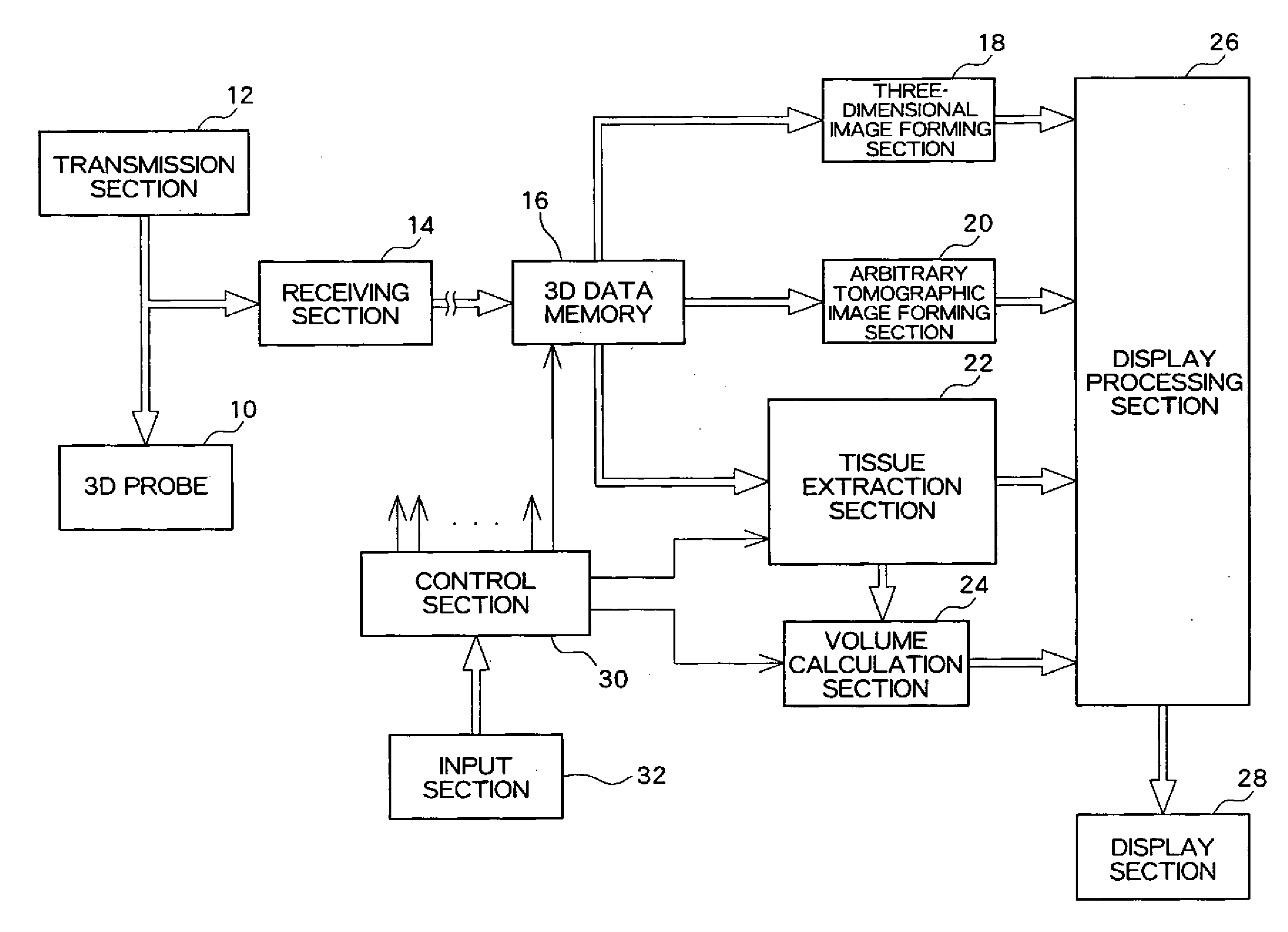
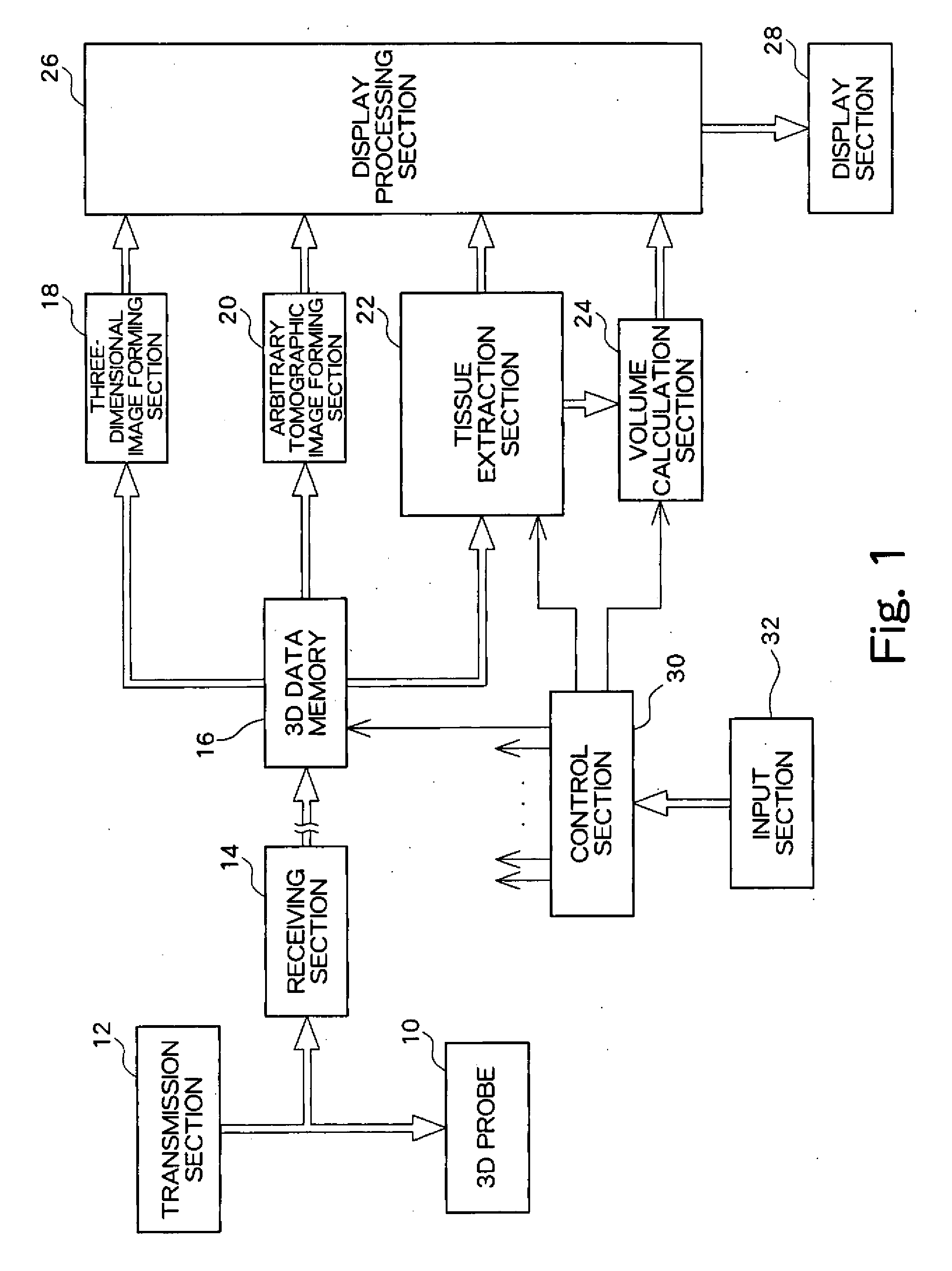
[0045]FIG. 1 is a block diagram showing the overall structure of an ultrasound diagnosis apparatus according to an embodiment of the present invention. This ultrasound diagnosis apparatus is intended for use in the medical field, and, in particular, has a function of extracting an object tissue within a living body and calculating the volume thereof. The object tissue may include a placenta, a malignant tumor, a gallbladder, a thyroid gland, and so on. In the following examples, processing of extracting a placenta as an object tissue will be described.

[0046]Referring to FIG. 1, a 3D (three-dimensional) probe 10 is an ultrasound transmitter / receiver which is used in contact with a body surface or used in a state where it is inserted into cavities of humans. In this embodiment, the 3D probe 10 includes a 2D (two-dimensional) array transducer which is composed of...


PUM


Abstract

An ultrasound diagnosis apparatus capable of extracting an object tissue within a living body with high precision is provided. A row of reference cross sections is set with regard to volume data. The row of reference cross sections includes a plurality of manual tracing reference cross sections and the remaining sections, i.e. a plurality of automatic tracing reference cross sections. With regard to the manual tracing reference cross sections, manual tracing processing and automatic correction processing is applied. With regard to the automatic tracing reference cross sections, automatic generation processing and automatic correction processing for interpolation tracing lines is applied. Finally, the object tissue is extracted or the volume thereof is calculated based on a plurality of tracing lines. When the similarity between shapes of adjacent manual tracing lines is small, one or a plurality of manual tracing lines may be additionally provided between these tracing lines.

Description

BACKGROUND[0001]1. Technical Field[0002]The present invention relates to an ultrasound diagnostic apparatus and a volume data processing method, and more particularly to specification or measurement of an object tissue within a three-dimensional space.[0003]2. Related Art[0004]A three-dimensional space (a three-dimensional data acquisition space) can be formed by scanning a scanning plane, which is formed by scanning an ultrasound beam, in the direction orthogonal to the scanning plane. A Disk Summation method is conventionally known as a method for extracting an object tissue existing within a three-dimensional space and calculating the volume thereof. With this conventional method, each of a plurality of slice data items which are aligned across the object tissue is displayed as a tomographic image, and the outline of the object tissue is manually traced on each tomographic image, enabling the cross sectional area in each slice to be calculated. Then, the cross sectional area and ...

Claims


Application Information

Patent Timeline
IPC IPC(8): A61B8/00
CPCA61B8/14A61B8/483G06T7/0083G06T2207/30004G06T2207/10136G06T2207/20104G06T7/602G06T7/12G06T7/62
Inventor MURASHITA, MASARUNAKAMURA, MASASHI
Owner FUJIFILM HEALTHCARE CORP
Why Eureka
Try Eureka