


Quick Research
Generate reliable direction feasibility study reports for your R&D in just a few steps.

Technical Q&A
Discover and master advanced knowledge NOW. Basics, ideas, possibilities, all at once.

Find Solutions
As an expert in R&D theories, this can generate solutions to your technical problems instantly.

Evaluate Feasibility
Analyze your overall solution with one click, know your potential R&D risks in advance.

Monitor Landscape
Get weekly tech updates, stay abreast of the latest tech innovations and key insights.
X-Ray Visible Drug Delivery Device
a drug delivery device and x-ray technology, applied in the direction of drugs, sexual disorders, prosthesis, etc., can solve the problems of difficult removal of implants, difficult to visualize by mri, and relatively complicated and expensive problems
- Summary
- Abstract
- Description
- Claims
- Application Information
AI Technical Summary
Benefits of technology
Problems solved by technology
Method used
Image

Examples
example 1
Preparation of Two-Layered Implant Containing Barium Sulphate in the Core
[0046]Preparation of two layered implant containing barium sulphate in the core consisted of two steps, i.e. manufacturing of core granulate (pre-mixing and blend extrusion) containing a mixture of etonogestrel (3-keto desogestrel), barium sulphate and EVA-28 copolymer and manufacturing of a co-axial fiber consisting of the core and a skin layer of EVA-14 copolymer.
[0047]The core material was prepared by adding the desired amount (e.g. 52.5 wt % etonogestrel, 36 wt % EVA, 11.5 wt % Barium sulphate) of ingredients to a stainless steel drum after which the powder mixture was pre-mixed by rotating the drum on a rhönrad, or equivalent, at 47 rpm. The powder mixture was subsequently fed to a Berstorff ZE25 co-rotating twin screw extruder (or equivalent) and blend extruded at an extrusion temperature of 125° C. Blend extrusion resulted in strands in which etonogestrel (3-keto desogestrel) and barium sulphate were hom...
example 2
Preparation of Two Layered Implant Containing Barium Sulphate in the Skin
[0049]Preparation of two layered implant containing barium sulphate in the skin consisted of three steps, i.e. manufacturing of core granulate (pre-mixing and blend extrusion) containing a mixture of etonogestrel (3-keto desogestrel) and EVA-28 copolymer, manufacturing of skin granulate (pre-mixing and blend extrusion) containing a mixture of Barium sulphate and EVA-14 copolymer, and manufacturing of a co-axial fiber consisting of the core and a skin layer.
[0050]The core material (e.g 60 wt % etonogestrel and 40 wt % EVA-28) and skin material (e.g. 20 wt % barium sulphate and 80 wt % EVA-14) were prepared by adding the desired ingredients to a stainless steel drum after which the powder mixtures were premixed by rotating the drum on a rhönrad, or equivalent, at 47 rpm.
[0051]The core powder mixture was subsequently fed to a Berstorff ZE25 co-rotating twin screw extruder (or equivalent) and blend extruded at an e...
example 3
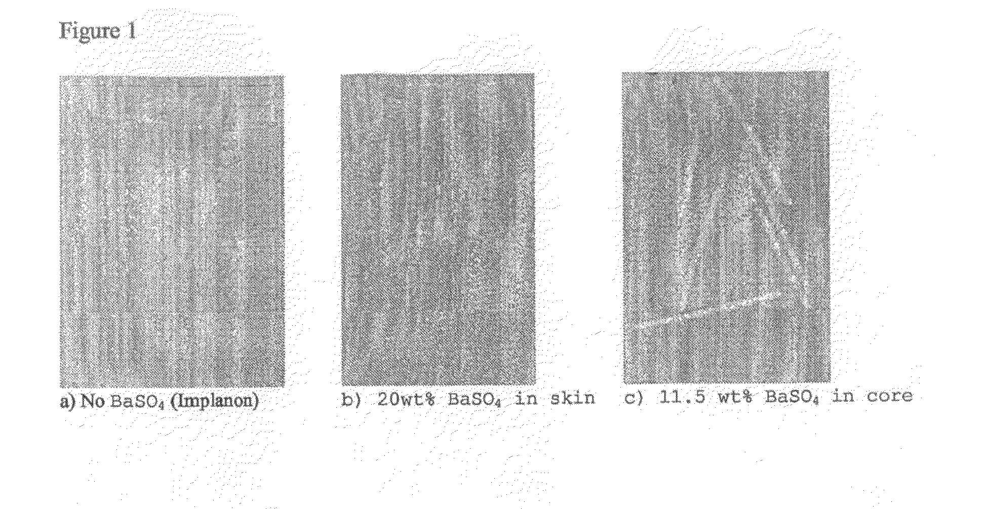
Comparison of X-Ray Visibility Between Implant Containing Barium Sulphate in the Core, Implant Containing Barium Sulphate in the Skin and Implant without Barium Sulphate (Implanon)
[0053]X-ray photographs were taken from implants and subsequently the X-ray visibility between implants having barium sulphate in either core or skin versus x-ray visibility of implants without barium sulphate (Implanon) were compared. FIG. 1 demonstrates that incorporation of barium sulphate in the skin layer hardly improved the x-ray visibility when compared to implants without barium sulphate. However, incorporation of barium sulphate into the core greatly improved the X-ray visibility of the implant.
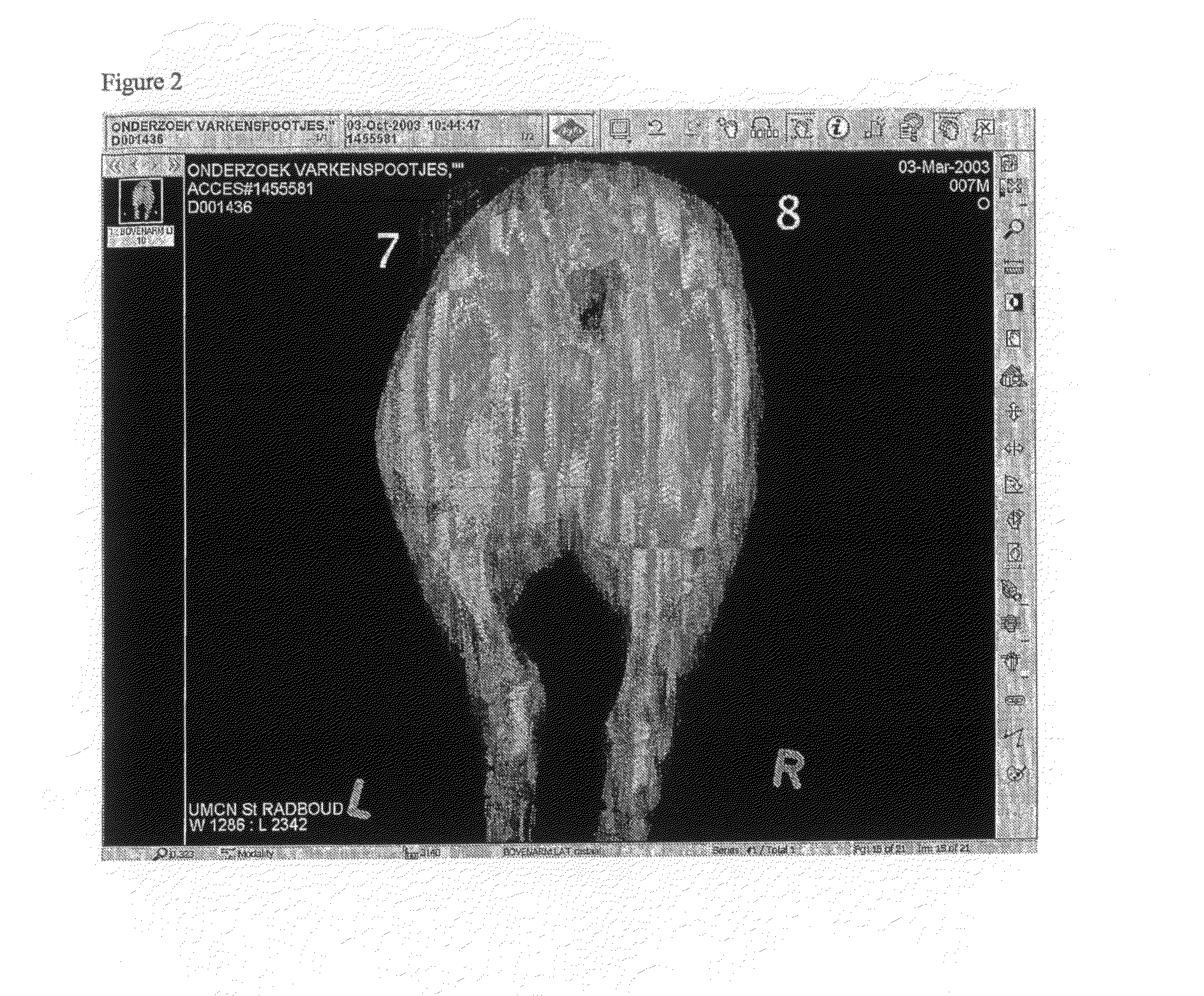
[0054]The x-ray visibility of the implant with barium sulphate in the core was also tested in vivo in pig tissue. For this purpose implants having barium sulphate in the core and implants without barium sulphate (Implanon) were inserted in hind legs of pigs and subsequently X-ray photographs were taken. FIG...
PUM
 Abstract
Description
Claims
Application Information
 - R&D Engineer
- R&D Manager
- IP Professional
- Industry Leading Data Capabilities
- Powerful AI technology
- Patent DNA Extraction
Browse by: Latest US Patents, China's latest patents, Technical Efficacy Thesaurus, Application Domain, Technology Topic, Popular Technical Reports.
© 2024 PatSnap. All rights reserved.Legal|Privacy policy|Modern Slavery Act Transparency Statement|Sitemap|About US| Contact US: help@patsnap.com