Method and imaging system to compensate for patient movements when recording a series of medical images

Inactive Publication Date: 2008-04-10
SIEMENS AG
View PDF14 Cites 9 Cited by
  • Summary
  • Abstract
  • Description
  • Claims
  • Application Information

AI Technical Summary

Benefits of technology

[0012] Using this prior art as a starting point, the object of the present invention is to specify a method as well as an associated imaging system to compensate for the motion of patients, especially in the abdominal area, when recording a series of images in medical imaging, with which the motion of the patient can be compensated for while the images are being recorded without time-consuming user interaction, with the method being able to be implemented in realtime. The method and the associated imaging system should in particular improve image results in a digital subtraction angiography and roadmapping with the lowest possible outlay as regards the operator's time.
[0014] In the present method for compensating for patient motion when recording a series of images in medical imaging, in which a number of images of the area under examination of a patient are recorded at intervals with an imaging system and are related to one another, a plurality of breath-triggered mask images are recorded during a breathing cycle. With the same triggering filling images are recorded and mask images and filling images which are recorded for the same breathing states are subtracted from each other in realtime and the difference images are displayed in realtime. A much better match between the current filling image and the mask image is achieved in this way. Depending on the application between 2 and 40 mask images are recorded in one breathing cycle. The subtraction of specific mask images from filling images taken in an identical breathing state is a live process in one embodiment of the present invention, i.e. during the recording of the filling images, so that especially in the pathfinder method for example a secure catheter guidance is possible. Mask images and filling images and their difference are stored in the image processing system.
[0016] The present method, by contrast with most previously known methods of motion correction, manages without interaction with the operator. The proposed method only requires a breath sensor to be accommodated on the patient where necessary. Thereafter no further user interaction is required for motion compensation. The principle of previous methods for retrospective image processing dictates that they only operate approximately. It is barely possible to correct large movements using these methods, and small movements can only be approximately corrected. The proposed method operates with high precision even for large movements, so that the need to record a mask image more than once is avoided. The saves time, contrast means and reduces the applied x-ray dose in the case of x-ray image recording. The present method also makes it possible in particular cases to dispense with sedating or anaesthetizing the patient merely for the purpose of minimizing motion artefacts.
[0017] Naturally, with the present method, after the at least approximate compensation of the movement, retrospective image processing methods can also be used in order to further improve the image results. The approximate compensation by modifying the geometrical circumstances of the imaging system can be used in this case to compensate for rough movements, while remaining small errors can be rectified by retrospective image processing.

Problems solved by technology

With longer interventions it is not possible to get the patient to hold their breath for so long, for example, when a catheter is being set.

Method used


Image

  • Method and imaging system to compensate for patient movements when recording a series of medical images
  • Method and imaging system to compensate for patient movements when recording a series of medical images

Examples


Embodiment Construction

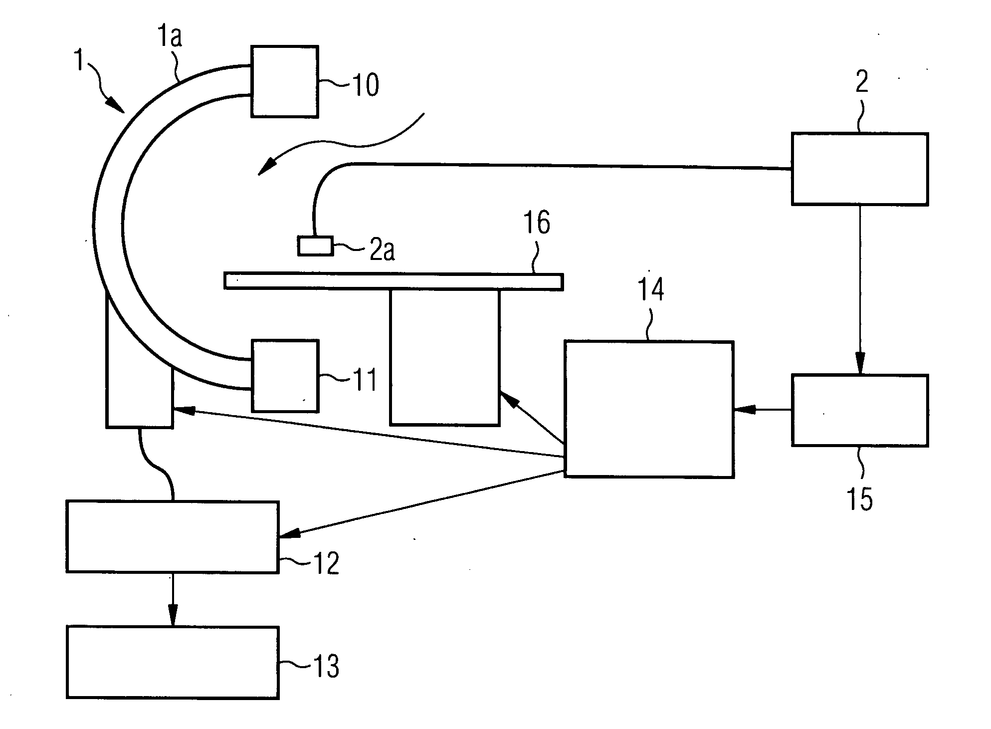
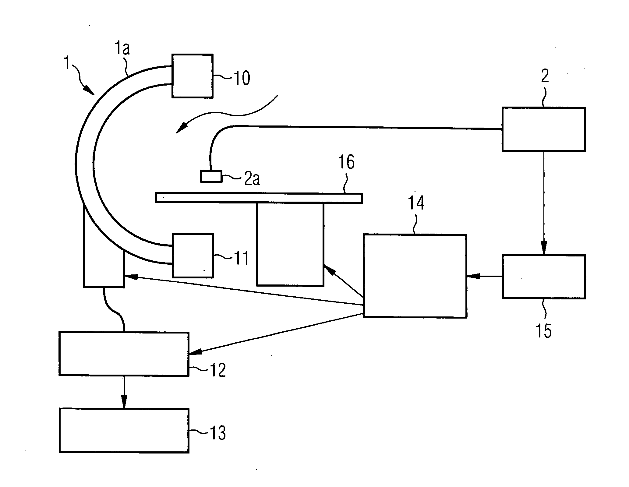
[0020] The present method is described below with reference to an x-ray angiography system for applications in neuro-radiology. The method can naturally also be used in other fields in which digital subtraction angiography and / or roadmapping are employed. The present method can also be used with other medical imaging techniques involving having to record a series of images and relate them to one another.

[0021] An x-ray angiography system 1 for neuro-radiology, which is shown schematically in FIG. 1, is used to record the images. The x-ray angiography system 1 includes a C-arm 1a which can be rotated around two axes, to which an x-ray tube 10 and a detector 11 arranged opposite the x-ray tube are attached, an image processing unit 12 and an image display unit 13. Furthermore this system includes the motor-driven adjustable patient table 16, a control unit 14 for image recording control as well as the compensation unit 15. Rotation of the C-arm 1a allows different projections of the ...


PUM


Abstract

The present invention relates to a method and imaging system to compensate for patient movements when recording a series of medical images, in which a number of images of an area of a patient to be examined are recorded at intervals and are related to one another, especially for movement compensation in Digital Subtraction Angiography (DSA) or roadmapping (RDMP) in the abdominal area. A plurality of breath-triggered mask images is recorded during a breathing cycle. Frilling images are recorded with the same triggering and mask images and filling images which are recorded for the same breathing states are subtracted from each other in realtime and the difference recordings are displayed in realtime.

Description

CROSS REFERENCE TO RELATED APPLICATIONS [0001] This application claims priority of German application No. 10 2006 047 719.7 filed Oct. 09, 2006, which is incorporated by reference herein in its entirety. FIELD OF THE INVENTION [0002] The present invention relates to a method to compensate for patient movements when recording a series of medical images in which a number of images of an area under examination of the patient are taken at intervals with an imaging system and are compared to one another, as is the case for example in Digital Subtraction Angiography (DSA) or roadmapping. The invention further relates to a medical imaging system with radiation source, detector, patient bed, image processing unit and image display unit which is embodied to execute the method. BACKGROUND OF THE INVENTION [0003] In a main area of application of the present method, the area of digital subtraction angiography, blood vessels of the human body are recorded with an imaging system, in this case an ...

Claims


Application Information

Patent Timeline
IPC IPC(8): G06K9/00
CPCA61B5/08A61B6/481A61B6/488A61B6/504A61B2019/5289A61B19/52A61B2017/00699A61B2019/5238A61B6/541A61B90/36A61B2090/364A61B2090/376
Inventor HILDEBRANDT, PETERPFLAUM, MICHAEL
Owner SIEMENS AG
Why Eureka
Try Eureka