Method and apparatus for combined gamma/x-ray imaging in stereotactic biopsy
- Summary
- Abstract
- Description
- Claims
- Application Information
AI Technical Summary
Benefits of technology
Problems solved by technology
Method used
Image
Examples
specific example 1
[0095]This example utilized an about 5 cm thick plastic breast phantom with an about 6 mm hollow sphere lesion located near the center of the breast phantom. The breast volume was filled with a Tc99m solution with a concentration of about 0.33 μCi / ml and the lesion volume concentration was about 1.98 μCi / ml. Two opposing 10 minute acquisitions were obtained. A pair of Co-57 point sources was taped to the edge of the phantom to aid in alignment of the opposing views.
[0096]The phantom was then emptied and refilled with F-18 for imaging with a dedicated small field-of-view positron breast imaging system, (PEM). The breast volume contained a concentration of about 0.08 μCi / ml and the lesion concentration was about 6:1 over that of the breast. Imaging was conducted for about 20 minutes and image reconstruction was completed using a classical back-projection tomography techniques.
[0097]FIG. 9A shows phantom images from the single gamma camera system, with the resulting images from the Tc9...
specific example 2
[0102]In the second experiment, about a 4.5 cm thick compressed breast phantom with a Tc99m concentration of 0.9 μCi / ml was prepared containing three lesions (two of about 8 mm diameter and one of about 6 mm diameter) containing an about 6:1 concentration over background and two 10 minute static acquisitions were obtained. A SPECT acquisition was performed with the same lesions and background solution transferred to a cylindrical (“uncompressed”) phantom with a diameter of about 9.25 cm. The SPECT acquisition angular sampling was set at about 3 degrees / step and the imaging time was set at about 30 seconds / frame. These parameters were selected to simulate about a 40 minute patient imaging time with a dual head system. Image reconstruction was obtained using a filtered back-projection technique.
[0103]In the planar imaging case, both the about 8 mm lesions and the about 6 mm lesion were visible in detector position 1 (Panel I of FIG. 10A). However, the about 6 mm lesion was not seen fr...
specific example 3
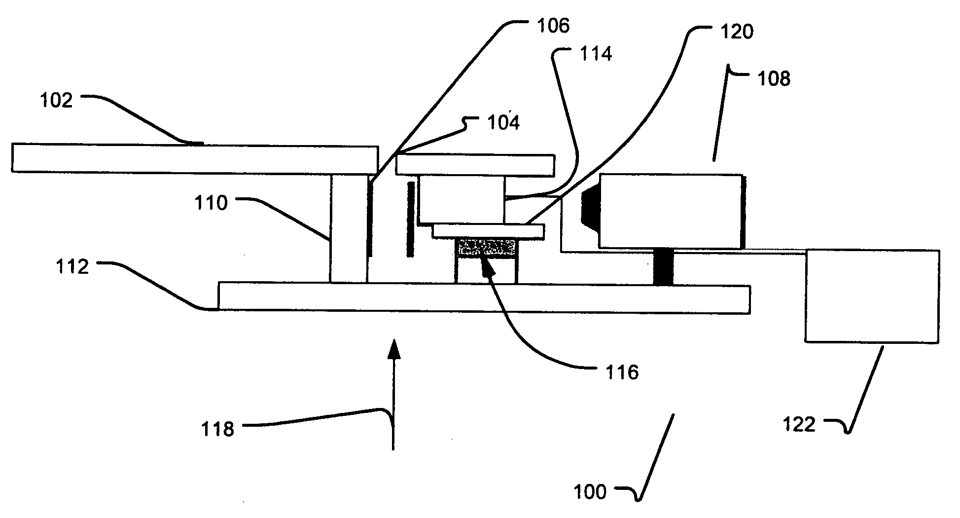
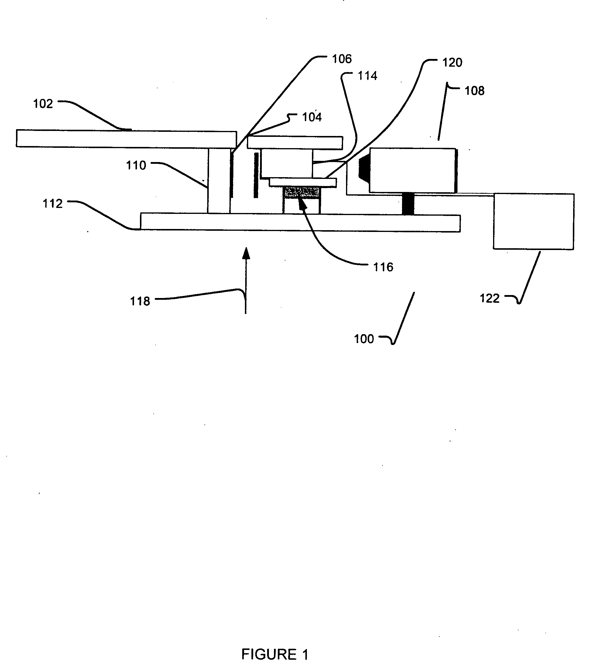
[0107]The patients enlisted in this study (N=55) were selected after a suspicious finding was reported in a routine X-ray screening mammogram. Using the mammographic films as guidance, the patients were placed on the stereotactic system table and the breast was compressed with a 5 cm×5 cm compression paddle (mean compression tissue thickness of about 5.96 cm, SD=about 1.41 cm). Scout views were obtained with the X-ray system until it was verified that the region-of-concern demonstrated in the mammogram was in the field-of-view. The mini gamma camera was then mounted to the X-ray system gantry in the needle driver position, see FIG. 1. A radiotracer was administered via venous puncture and an acquisition was initiated at the time of injection for about 10 minutes. Digital X-ray images were stored as high-resolution tiff images, and about 10-minute static gamma camera images were obtained for all patients. Additionally, dynamic data was stored in list mode for 33 of the 55 cases and r...
PUM
 Abstract
Description
Claims
Application Information
 - R&D
- Intellectual Property
- Life Sciences
- Materials
- Tech Scout
- Unparalleled Data Quality
- Higher Quality Content
- 60% Fewer Hallucinations
Browse by: Latest US Patents, China's latest patents, Technical Efficacy Thesaurus, Application Domain, Technology Topic, Popular Technical Reports.
© 2025 PatSnap. All rights reserved.Legal|Privacy policy|Modern Slavery Act Transparency Statement|Sitemap|About US| Contact US: help@patsnap.com


