RNA-selective probes for live cell imaging of nuclear structure and function

a live cell and probe technology, applied in the field of live cell imaging probes of nuclear structure and function, can solve the problems of inconvenient availability of rna-specific dyes for staining live cells, inability to widely prove their usefulness, and inability to publish their molecular structur

Active Publication Date: 2008-03-13
NEW YORK UNIV
View PDF2 Cites 24 Cited by
  • Summary
  • Abstract
  • Description
  • Claims
  • Application Information

AI Technical Summary

Benefits of technology

[0010] A third aspect of the present invention relates to a second fluorescent compound, referred to as E144, having the formula:
[0011] Another aspect of the present invention relates to a process of making the second fluorescent compound having the formula:
[0012] Another aspect of the present invention includes a process of making a the third fluorescent compound, referred to as E36, having the formula:
[0013] Another aspect of the

Problems solved by technology

In contrast, RNA-specific dyes for staining live cells are not readily available.
M. T. Z. Spence, eds. pp 327, 710-711), but its usefulness has not been widely proven and its molecular structure has not yet been published.
However, finding RNA-selective compounds for live cell imaging is difficult, because small nucleic acid binding molecules generally have better affinity to double-stranded DNA compared to single-stranded RNA.
In addition, the living cell system is complicated by the fact that it is rich in proteins and membranes that may lead to non-specific binding of hydrophobic imaging probes.

Method used


Image

  • RNA-selective probes for live cell imaging of nuclear structure and function
  • RNA-selective probes for live cell imaging of nuclear structure and function
  • RNA-selective probes for live cell imaging of nuclear structure and function

Examples


example 1

Materials and Instruments

[0039] Unless otherwise noted, chemical materials and solvents were purchased from commercial suppliers (Sigma-Aldrich, St. Louis, Mo. and Acros, Pittsburgh, Pa.) and were used without further purification. RNA (type VI from torula yeast, Sigma) was used for primary screening. RNA, 16S- and 23S-ribosomal from E. coli, (Roche, Indianapolis, Ind.) and DNA from salmon testes (Sigma) were used for dye concentration dependency study. TE buffer (Tris-EDTA sterile solution at pH 7.5, nuclease and protease-free 0.2 μm filtered solution, GE, Piscataway, N.J.) was used in all solution experiments. A Gemini XS fluorescent plate reader was used in obtaining the excitation and emission spectra in the primary screening and titration assay studies. Greiner 96 or 384 well polypropylene black plates were used in the experiments. Full spectra data of the selected three dyes were obtained by Hitachi F-2500 fluorophotometer. Absorbance spectra were measured using SpectraMax Pl...

example 2

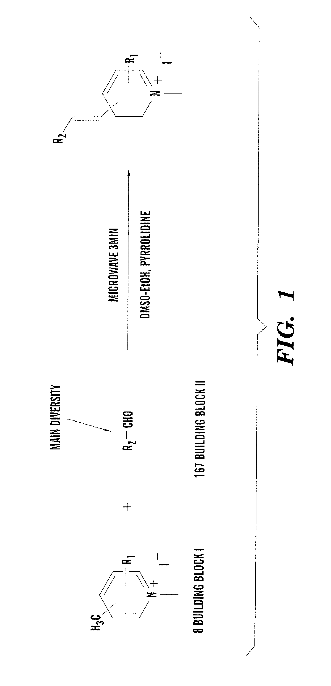
General Procedure for Synthesis of Building Block I

[0040] The pyridine derivative (2.04 mmol) and iodomethane (2.14 mmol) in ethyl acetate were refluxed overnight. After it was cooled to room temperature, the crystallized methylated products were filtered and washed with ethyl acetate three times and dried in vacuum.

example 3

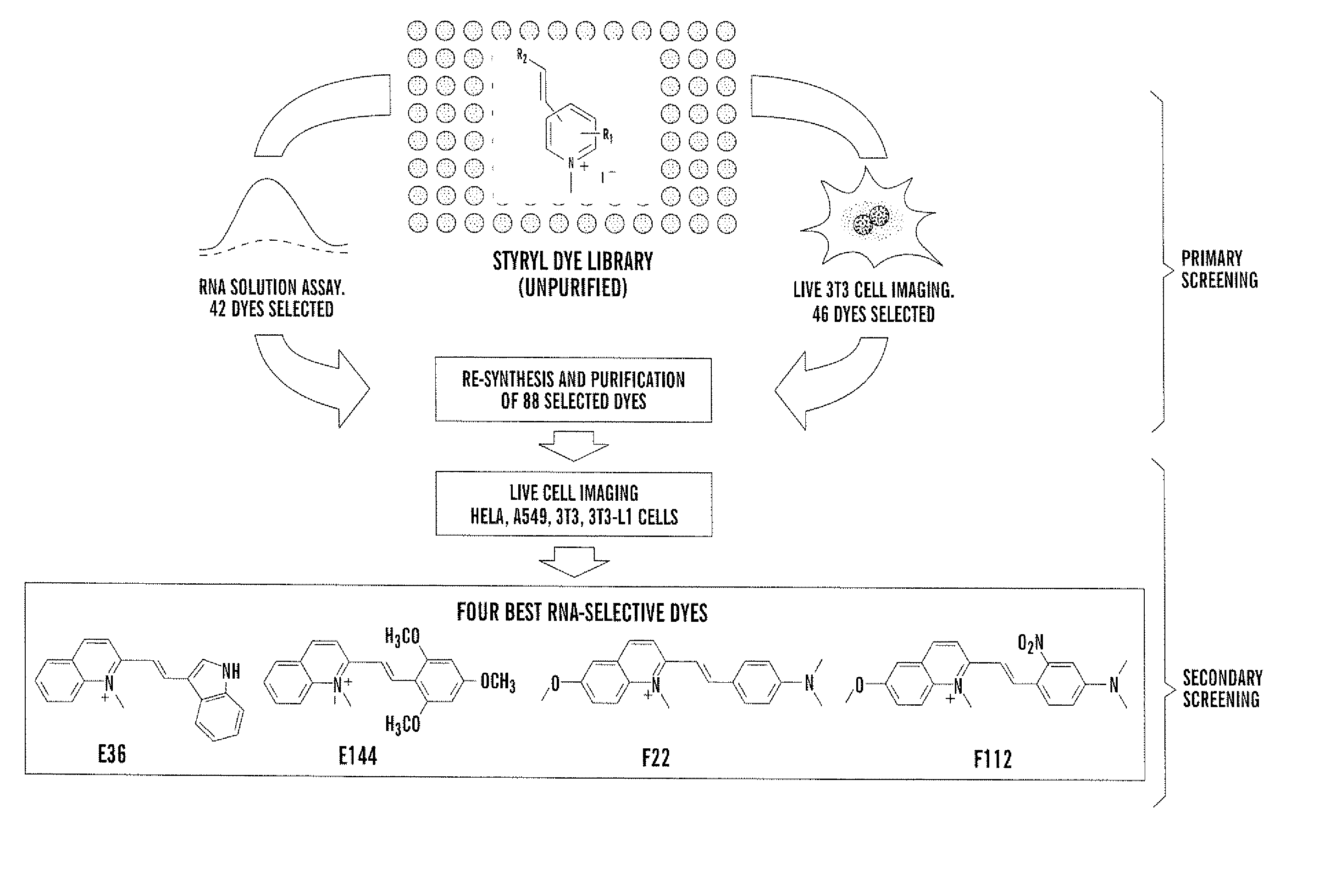
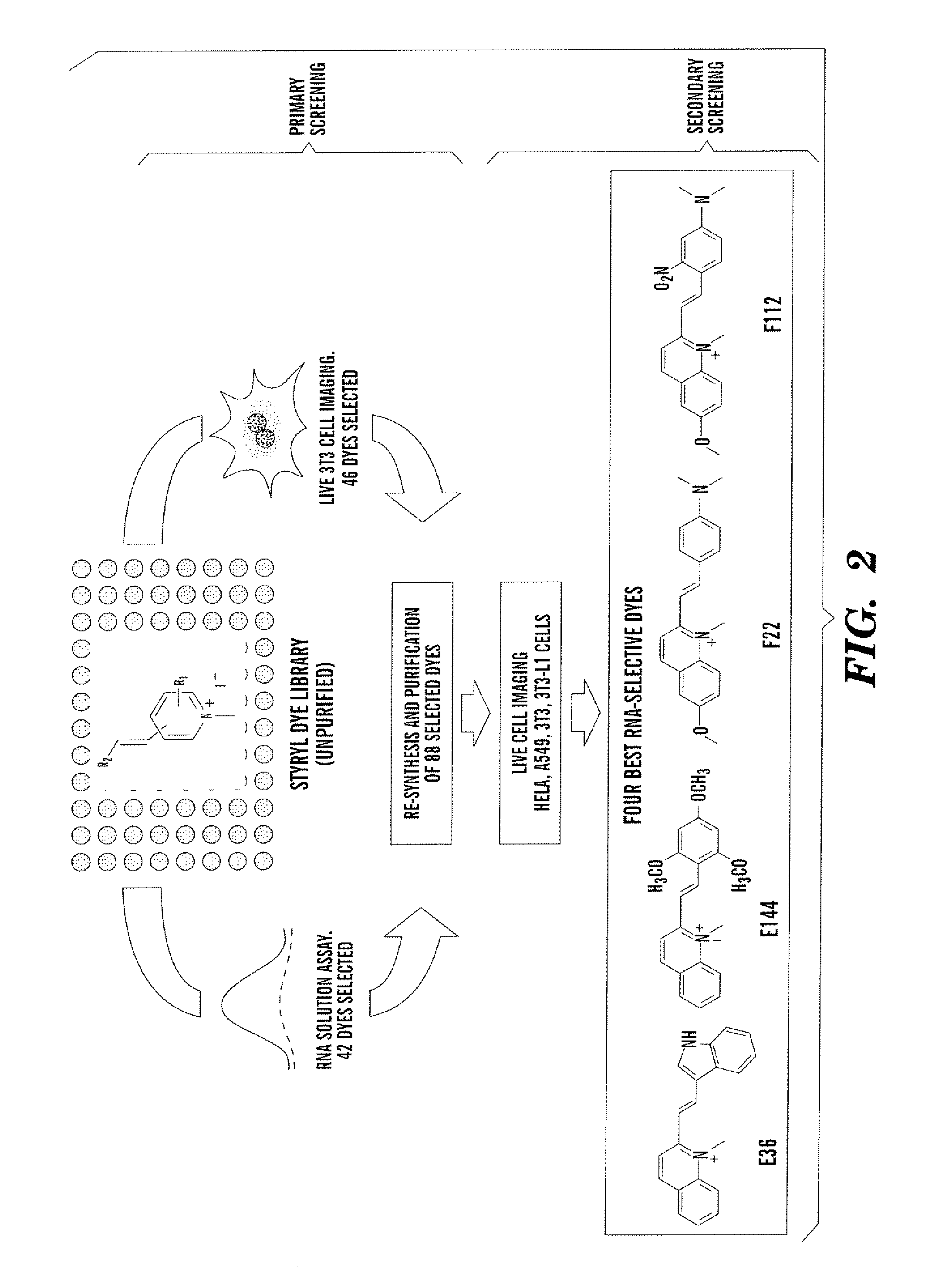
General Procedure for Synthesis of Styryl Dye Library

[0041] Building blocks I and II were dissolved separately in absolute ethanol (100 mM) as stock solutions. In 96-well Gemini polypropylene microtiter plates, mM of each reactant, 30 μL DMSO, and 0.1 μL pyrrolidine were added using a multi-pipette. The condensation reaction was carried out in an 800 watt microwave, three minutes for each plate. Product yield and purity were identified by LCMS.


PUM

PropertyMeasurementUnit
Lengthaaaaaaaaaa
Lengthaaaaaaaaaa
Lengthaaaaaaaaaa

Abstract

The present invention is directed to fluorescent compounds and methods of making said compounds that selectively bind to cellular RNA. The fluorescent compounds of the present invention are useful for live cell imaging applications.

Description

[0001] This application claims the benefit of U.S. Provisional Patent Application Ser. No. 60 / 780,779, filed Mar. 9, 2006.[0002] The subject matter of this application was made with support from the United States Government under the National Science Foundation, Grant No. CHE-0449139, the National Institutes of Health, Grant No. RO1-GM078200, the National Center for Research Resources / National Institutes of Health, Grant No. C06 RR-16572. The U.S. Government may have certain rights.FIELD OF THE INVENTION [0003] The present invention is directed to probes for live cell imaging of nuclear structure and function. BACKGROUND OF THE INVENTION [0004] Live cell imaging using fluorescent microscopy technology has allowed investigation of the transcriptional dynamics of the cell nucleus (Tsien et al., “Seeing the Machinery of Live Cells”, Science 280:1954-1955 (1998); Stephens et al., “Light Microscopy Techniques for Live Cell Imaging,”Science 300.82-86 (2003)). RNA molecules in living cells...

Claims


Application Information

Patent Timeline
IPC IPC(8): C12Q1/68C07D215/04C07D215/12C07D215/20C12Q1/02
CPCC07D215/10C07D215/14C07D215/20C07D401/06C12Q1/68C12Q1/6841G01N33/5079G01N33/5005G01N33/5076C12Q2563/173
Inventor CHANG, YOUNG-TAELI, QIANROSANIA, GUSTAVO
Owner NEW YORK UNIV
Why Eureka
Try Eureka