
Device for cartilage repair

a cartilage repair and cartilage technology, applied in the field of cartilage repair devices, can solve the problems of severe donor limitations, inability to consistently demonstrate successful repair of articular lesions, and inability to achieve consistent results of micro-fracture surgery

Inactive Publication Date: 2008-02-21
MISHRA ALLAN
View PDF5 Cites 72 Cited by
  • Summary
  • Abstract
  • Description
  • Claims
  • Application Information

AI Technical Summary

Problems solved by technology

Most of these methods have been shown to have some short term benefit in reducing symptoms (months to a few years) while none have been able to consistently demonstrate successful repair of articular lesions after the first few years.
Pharmacological modulation supplying growth factors to defect sites can augment natural repair but to date insufficiently so (Hunziker and Rosenberg, 1996; Sellers et al., 1997).
Allograft and autograft osteochondral tissue transplants containing viable chondrocytes can effect a more successful repair but suffer from severe donor limitations (Mahomed et al., 1992; Outerbridge et al., 1995).
Unfortunately, micro-fracture surgery is only inconsistently efficacious.
If the fill of the defect is inadequate, the clinical results are typically either equivocal or even poor.

Method used


Image

  • Device for cartilage repair
  • Device for cartilage repair
  • Device for cartilage repair

Examples


example 1

[0061]PRP was prepared using a centrifuge unit made by Harvest (Plymouth, Mass.). (Similar units are available as The Biomet GPS system, the Depuy Symphony machine and the Medtronic Magellan machine.) Approximately 55 cc of blood was drawn from the patient using a standard sterile syringe, combined with 5 cc of a citrate dextrose solution for anticoagulation, and then spun down to isolate the platelets according to the manufacturer's protocol. These platelets were then resuspended in approximately 3 cc of plasma. The resulting platelet rich plasma solution (PRP) was quite acidic and was neutralized with using approximately 0.05 cc of an 8.4% sodium bicarbonate buffer per cc of PRP under sterile conditions to approximately physiologic pH of 7.4. The PRP was not activated through addition of exogenous activators. This PRP composition is referred to herein as autologous platelet extract (APEX).

example 2

[0062]Fifty cc of whole blood is drawn from a patient, and then prepared according to the method of Knighton, U.S. Pat. No. 5,165,938, column 3. The PRP is activated according to Knighton using recombinant human thrombin. The degranulated platelets are spun down and the releasate containing supernatant is recovered. The releasate may be optionally pH adjusted to a pH of 7.4 using sodium bicarbonate buffer.

example 3

[0063]Thirty ml of whole blood were drawn from a patient. A platelet composition was prepared according to Example 1 of U.S. Pat. No. 5,510,102 to Cochrum, incorporated herein by reference in its entirety, except that no alginate is added to the platelet composition.


PUM


Abstract

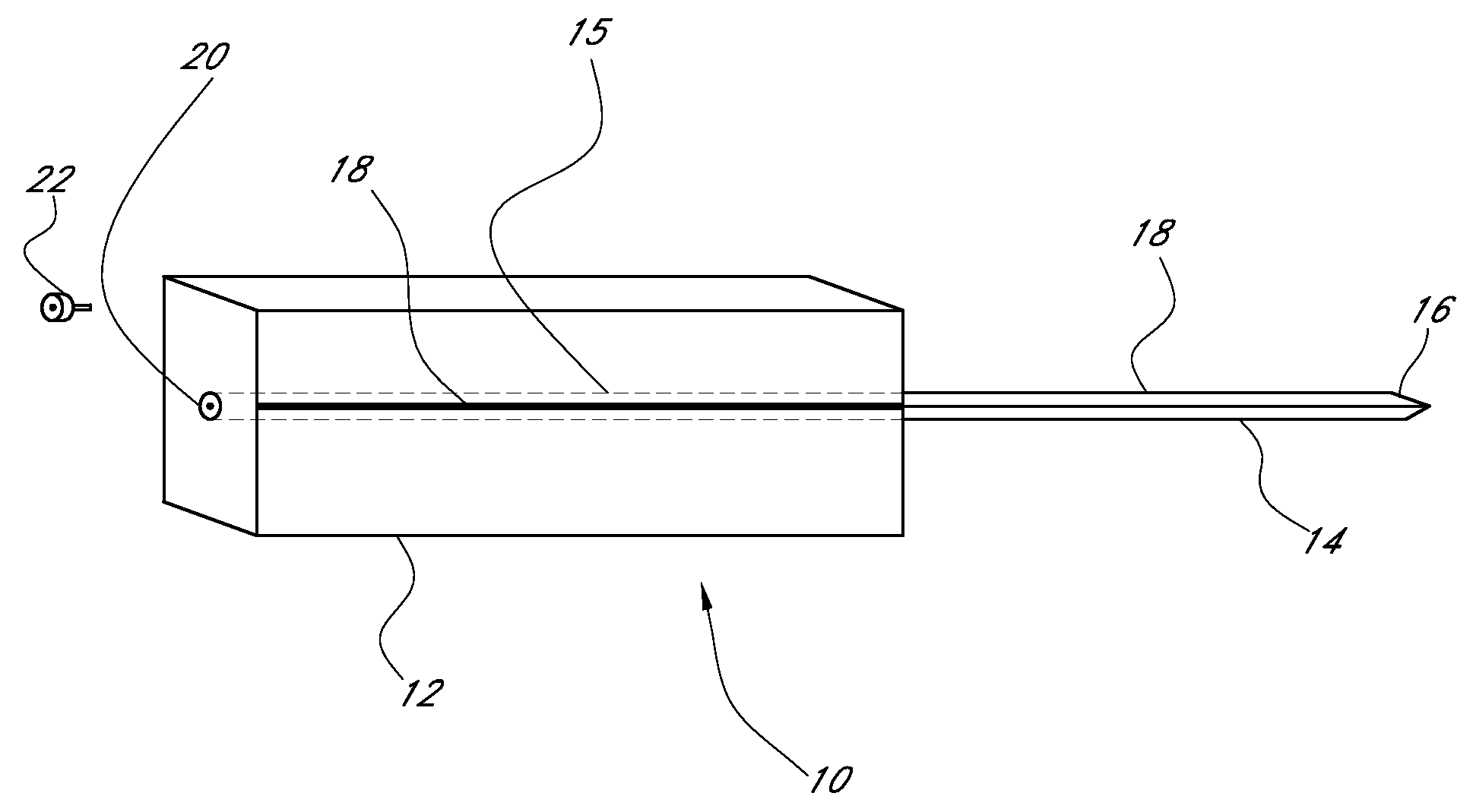
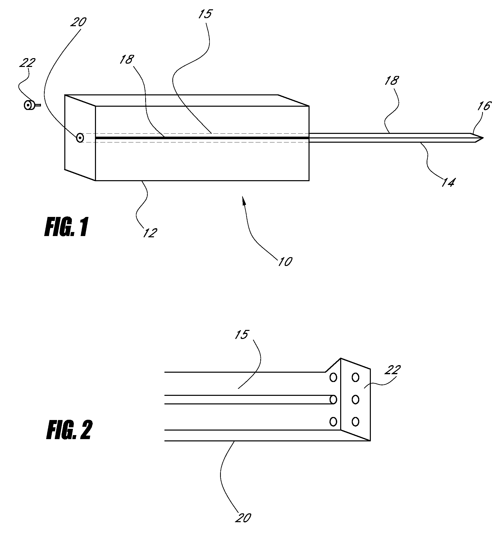
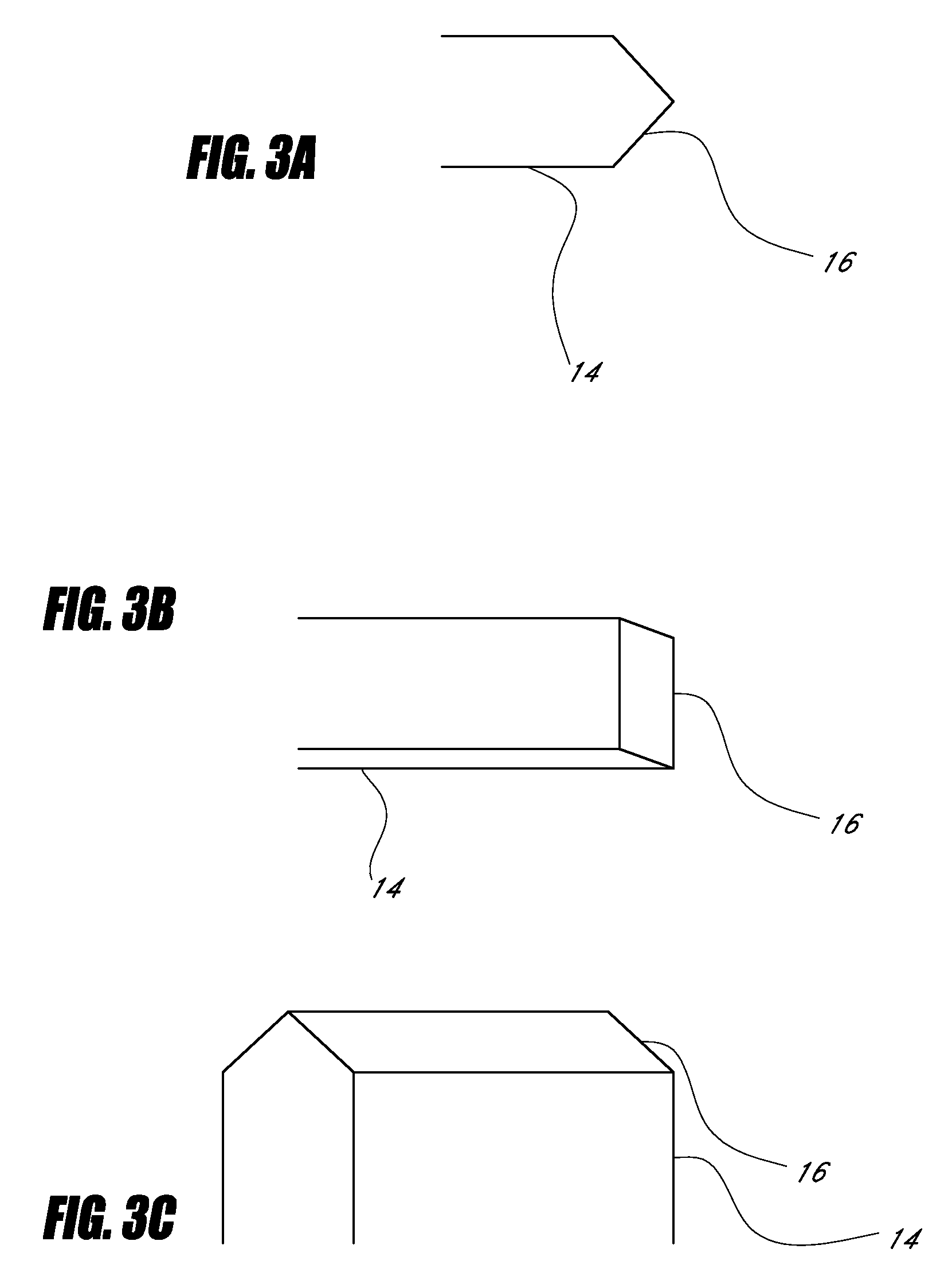
A surgical awl is disclosed for tissue repair. The surgical awl includes an internal lumen through which bioactive substances, such as platelet-rich plasma, may be delivered to a site of tissue or organ disease or dysfunction. In particular, the surgical awl is useful as a microfracture awl for cartilage repair augmented by delivery of platelet-rich plasma through the lumen of the device.

Description

CROSS-REFERENCE TO RELATED APPLICATIONS[0001]This application claims the benefit under 35 U.S.C. § 119(e) of U.S. Provisional Patent Application No. 60 / 838,064, filed Aug. 16, 2006, entitled “Device for Cartilage Repair,” which is hereby incorporated by reference herein in its entirety.BACKGROUND[0002]1. Field of the Invention[0003]The invention relates to a device for cartilage repair and methods of using the device to repair cartilage and to deliver bioactive substances such as platelet-rich plasma to tissues.[0004]2. Description of the Related Art[0005]Current clinical treatments for symptomatic cartilage defects involve techniques aimed at: 1) removing surface irregularities by shaving and debridement 2) penetration of subchondral bone by drilling, fracturing or abrasion to augment the natural repair response 3) joint realignment or osteotomy to use remaining cartilage for articulation 4) pharmacological modulation 5) tissue transplantation and 6) cell transplantation (Newman, 1...

Claims


Application Information

Patent Timeline
Patent Type & Authority Applications(United States)
IPC IPC(8): A61B17/56A61B17/32
CPCA61B17/1604A61B17/3468A61B17/7061A61B17/3478A61B17/3472
Inventor MISHRA, ALLAN
Owner MISHRA ALLAN
Learn More