
Methods and devices for analyte detection

a technology of analyte detection and medical devices, applied in the field of medical devices and kits for analyte detection, can solve the problems of inconvenient, expensive, or other deficiencies of current assay protocols, and the extensive handling of the components of the technique,

Active Publication Date: 2008-01-10
PROTEINSIMPLE
View PDF62 Cites 22 Cited by
  • Summary
  • Abstract
  • Description
  • Claims
  • Application Information

AI Technical Summary

Benefits of technology

[0007] The present invention provides methods, devices, and kits for detecting one or more analytes of interest in a sample. In some embodiments, methods of detecting at least one analyte in a sample are provided, characterized in that: one or more analy

Problems solved by technology

However, currently available assay protocols are inconvenient, expensive, or have other deficiencies.
First, as the above description makes clear, the processing is very complex.
Second, there is extensive handling of the components of the technique.
The components can be damaged during this handling, in particular the fragile sheet of gel.
Third, it takes a considerable amount of time to arrive at just a single blot.
During the beginning of the process the accuracy of the technique is affected by migration of the proteins until they are immobilized in the blotting material, which can result in band broadening.
Fourth, the variability introduced by the complexity of the handling and processing can require the process to be repeated several times before acceptable results are obtained.
Fifth, the variability in the results often requires subjective decisions to be made in reading the results of the blot.
This subjectivity reduces the ability to obtain quantifiable, objective results and frequently limits the technique to practice by highly trained and experienced personnel.
Sixth, the variability and complexity of the process impedes the ability to automate the process.
Seventh, the technique has low sensitivity and generally is only effective with the contents of hundreds of thousands or millions of cells.
Certainly, the technique cannot be used to analyze the enzymes of an individual mammalian cell.
Eighth, the quantitation of the technique is poor.
For one example, the agitation process may fail to cause the uniform binding of reporter substrates to the analytes in the blotting material.
Finally, the process can require large quantities of expensive probe and reporter antibodies to be used.
In sum, the Western gel blotting technique is generally complex, time-consuming, expensive, insensitive and inexact.

Method used


Image

  • Methods and devices for analyte detection
  • Methods and devices for analyte detection
  • Methods and devices for analyte detection

Examples


example 1

Fluorescence Detection of Green Fluorescent Protein (GFP)

[0175] Preparation of GFP sample for analysis: In a microcentrifuge tube, 40 μL of DI water, 1 μL of GFP at 1 mg / ml (Part number 632373, Beckton-Dickinson, San Jose, Calif., USA), 5 μL of bioPLUS pI 4-7 (Bio-World, Dublin, Ohio), and 2 μL of ATFB-PEG cross-linking agent (2 mM) were combined. The ATFB-PEG cross-linking agent consists of 15,000 MW branched polyethylene glycol (product number P4AM-15, SunBio, Anyang City, South Korea) in which each branch terminus was derivatized with an ATFB (4-azido-2,3,5,6-tetrafluorobenzoic acid) functionality (product number A-2252, Invitrogen Corporation, Carlsbad, Calif.).

[0176] Preparation of capillary: 100μ I.D.×375μ O.D. Teflon-coated fused silica capillary with interior vinyl coating (product number 0100CEL-01, Polymicro Technologies, Phoenix, Ariz.) was surface grafted on its interior with polyacrylamide containing 1 mole percent benzophenone. 4 cm sections of this capillary materia...

example 2

Chemiluminescence Detection of GFP

[0182] Preparation of GFP sample for analysis: In a microcentrifuge tube, 40 μL of DI water, 1 μL of GFP at 1 mg / ml (Part number 632373, BD Biosciences, San Jose, Calif., USA), 5 μL of bioPLUS pI 4-7 (Bio-World, Dublin, Ohio), and 2 μL of ATFB-PEG cross-linking agent (2 mM) were combined. The ATFB-PEG cross-linking agent consists of 15,000 MW branched polyethylene glycol (product number P4AM-15, SunBio, Anyang City, South Korea) in which each branch terminus was derivatized with an ATFB (4-azido-2,3,5,6-tetrafluorobenzoic acid) functionality (product number A-2252, Invitrogen Corporation, Carlsbad, Calif.).

[0183] Preparation of capillary: 100μ I.D.×375μ O.D. Teflon-coated fused silica capillary with interior vinyl coating (product number 0100CEL-01, Polymicro Technologies, Phoenix, Ariz.) was surface grafted on its interior with polyacrylamide containing 1 mole percent benzophenone. 4 cm sections of this capillary material were cleaved from longer...

example 3

Fluorescent Detection of Horse Myoglobin

[0189] Preparation of sample for analysis: In a microcentrifuge tube, 40 μL of DI water, 2 μL of 4 mg / ml purified horse myoglobin (Part number M-9267, Sigma-Aldrich, St. Louis, Mo., USA) myoglobin solution, 5 μL of Pharmalyte ampholyte pI 3-10 (Sigma, St. Louis), and 2 μL of ATFB-PEG cross-linking agent (2 mM) were combined. The ATFB-PEG cross-linking agent consists of 15,000 MW branched polyethylene glycol (product number P4AM-15, SunBio, Anyang City, South Korea) in which each branch terminus was derivatized with an ATFB (4-azido-2,3,5,6-tetrafluorobenzoic acid) functionality (product number A-2252, Invitrogen Corporation, Carlsbad, Calif.).

[0190] Preparation of capillary: 100μ I.D.×375μ O.D. Teflon-coated fused silica capillary with interior vinyl coating (product number 0100CEL-01, Polymicro Technologies, Phoenix, Ariz.) was surface grafted on its interior with polyacrylamide containing 1 mole percent benzophenone. 4 cm sections of this ...


PUM


Abstract

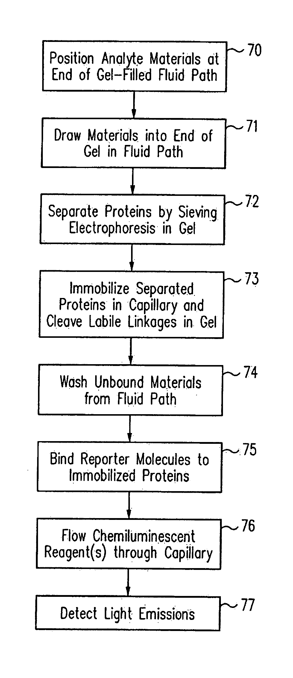
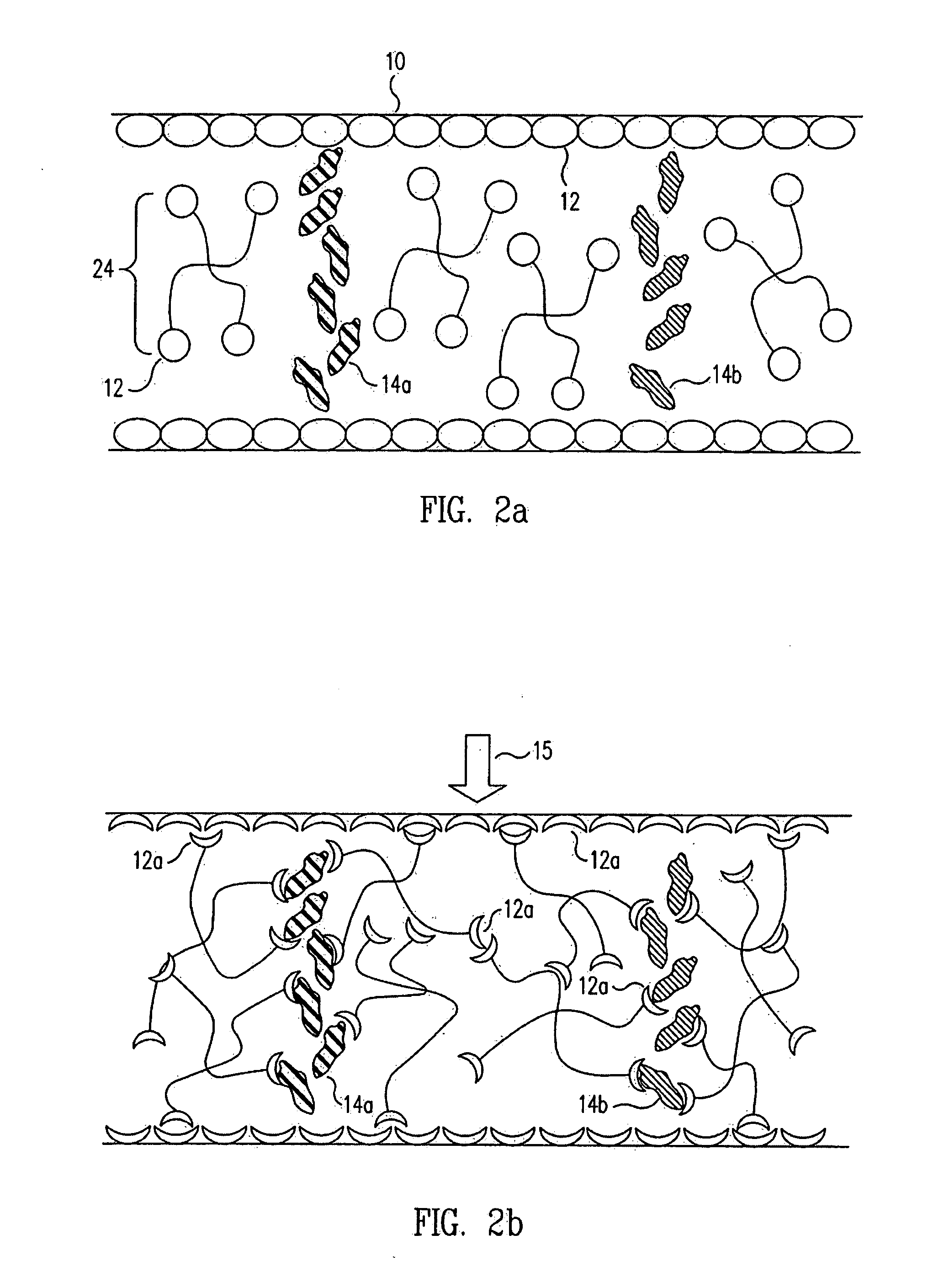
Methods and apparatus are provided to resolve analytes within a fluid path using isoelectric focusing, gel electrophoresis, or other separation means. Materials within the fluid path that are compatible with these separation means are used to attach resolved analytes to the wall of the fluid path. Attachment results from a triggerable event such as photoactivation, thermal activation, or chemical activation. In accordance with a further aspect of the present invention, the material in the capillary may also be disrupted, by either the triggerable event or a subsequent event such as melting or photocleavage. Thus, an open lumen or porous structure may be created within the fluid path, allowing unbound analyte materials to be washed from the fluid path, and detection agents to be washed into the fluid path. The separation-compatible materials may be polymerizable monomers, gels, entangled polymers or other materials.

Description

CROSS REFERENCES TO RELATED APPLICATIONS [0001] This application is a continuation-in-part of U.S. patent application Ser. No. 11 / 185,247, filed Jul. 19, 2005 and entitled “METHODS AND DEVICES FOR ANALYTE DETECTION,” which claims benefit under 35 U.S.C. §119(e) to application Ser. No. 60 / 589,139, entitled “Continuous Determination of Cellular Contents by Chemiluminescence,” filed Jul. 19, 2004 and application Ser. No. 60 / 617,362, entitled “Determination of Captured Cellular Contents,” filed Oct. 8, 2004, the disclosures of which are incorporated herein by reference in their entireties. This application further claims the benefit of U.S. provisional patent application Ser. No. 60 / 816,194, filed on Jun. 23, 2006, the disclosure of which is incorporated herein by reference in its entirety.FIELD OF INVENTION [0002] The present invention relates to medical devices and kits for analyte detection and various uses thereof. More particularly this invention relates to methods and devices for ...

Claims


Application Information

Patent Timeline
IPC IPC(8): G01N33/566B05D3/00G01N33/00
CPCB01L3/502715B01L3/50273B01L2200/0668B01L2400/0406B01L2400/0418G01N33/582B01L2400/0487G01N27/44726G01N27/44795G01N33/54366B01L2400/0421
Inventor O'NEILL, ROGER A.GLAZER, MARCYANG, TOM W.SUICH, DANIEL J.VOSS, KARL O.
Owner PROTEINSIMPLE
Learn More