
Anchoring and tethering system

Inactive Publication Date: 2007-11-15
AGA MEDICAL CORP MS US
View PDF99 Cites 349 Cited by
  • Summary
  • Abstract
  • Description
  • Claims
  • Application Information

AI Technical Summary

Benefits of technology

[0015] A first object of the present invention is to provide methods and devices for safe and effective transcatheter correction of the shape of heart chambers and / or valves for improved performance of the heart.

Problems solved by technology

Over time, disease and injury can cause damage to the heart.
In a diseased heart, the chambers can become swollen and distended leading to cardiac inefficiency and heart failure.
Such swelling can lead to damage to the electrical conduction paths in the heart that control its rhythm.
Likewise, the annulus of the mitral valve of the heart can become distended such that the leaflets do not fully close permitting blood to regurgitate (i.e., flow in the wrong direction through the valve).
Such regurgitation can result in inefficient pumping by the heart to a degree that is detrimental to patient health.
Such pharmaceuticals do not really remedy the condition, but instead can help control the effects of the condition or prevent a worsening of the condition.
Heart surgery typically entails great trauma to the patient and long recovery periods.
The risk of death during surgery is significant particularly because heart disease has often weakened the body, and particularly the heart, before such surgery is even attempted.
While the above-referenced patent applications disclose theoretical concepts for changing the size and shape of the annulus, those skilled in the art will recognize issues that make these proposed solutions impractical for general use.
Therefore, what affect changing the radius of curvature of the coronary sinus will have on the annulus of the mitral valve cannot be accurately predicted or easily controlled.
There is no teaching in these patent publications of how such precise alignment could be achieved.
Third, the coronary sinus only surrounds about half of the mitral valve.
Fourth, the devices shown could very well lead to significant occlusion of the coronary sinus which is an essential conduit for carrying blood.
While in theory, the system disclosed in the Starksen et al application might work, the system is impractical given the difficulty in aligning the device, setting the hook type anchors and applying proper tension between the anchors with the biasing means to achieve the proper shape all through a catheter.
There are risks of infection, damage to the muscle and thrombus formation between the various components that could lead to stroke or death.
The system shown in the Huynh et al is not practical for several reasons.
First, it would be extremely difficult to safely position the harpoon anchor in the coronary sinus, use it to puncture the coronary sinus, advance it across the atrium, and then securely and permanently fix it to the opposing heart wall.
Even if this could be accomplished, it would be difficult to ensure that the spot where this anchor is coupled to the wall and the point where it penetrates the coronary sinus will result in tension being supplied in a manner that corrects the shape of the annulus of the valve.
Likewise, the risk that the anchor could be pulled from the heart wall is significant.
Similar problems are inherent in the disclosure contained in U.S. Patent Publication No. 2005 / 0222488 (Change et al).
There is no guaranteeing that applying tension along that vector will result in proper reshaping of the annulus of the mitral valve.
However, the device shown is not implanted using a catheter.

Method used


Image

  • Anchoring and tethering system
  • Anchoring and tethering system
  • Anchoring and tethering system

Examples


Embodiment Construction

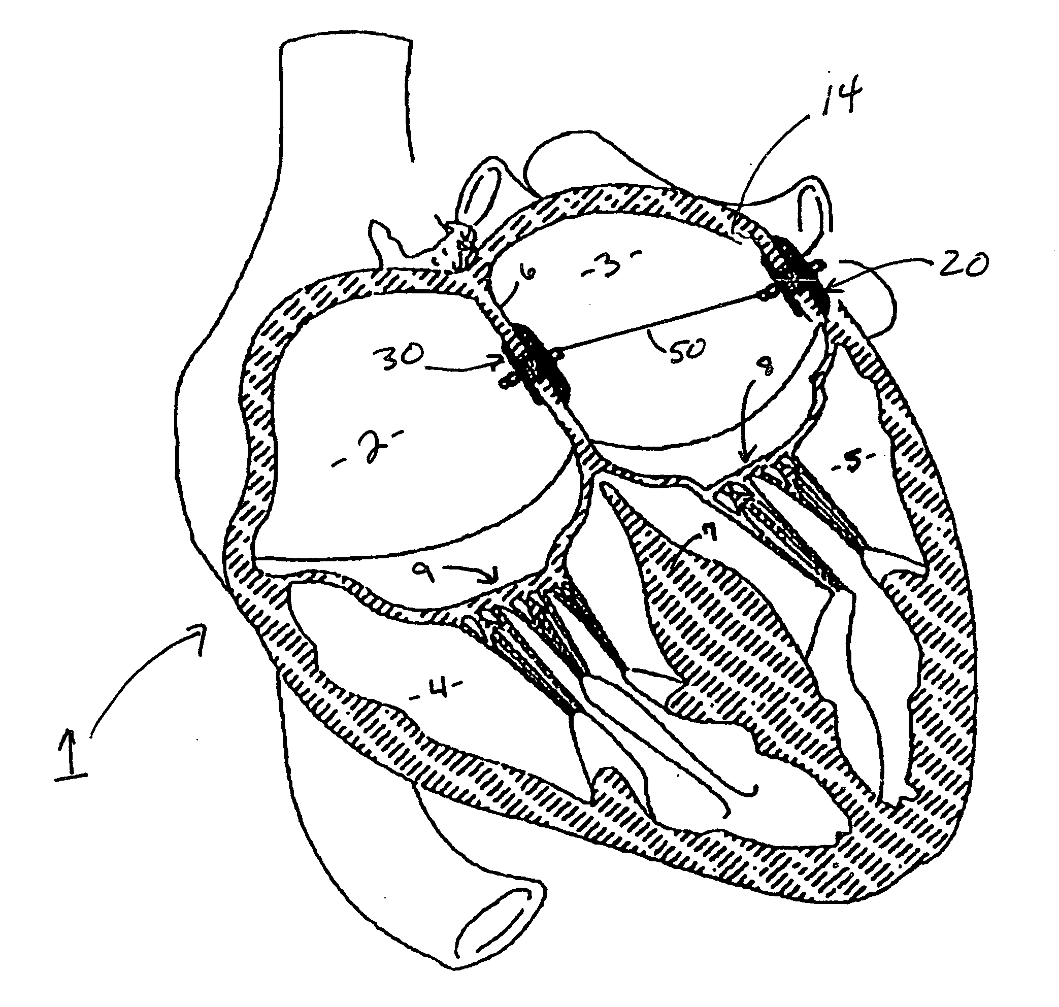
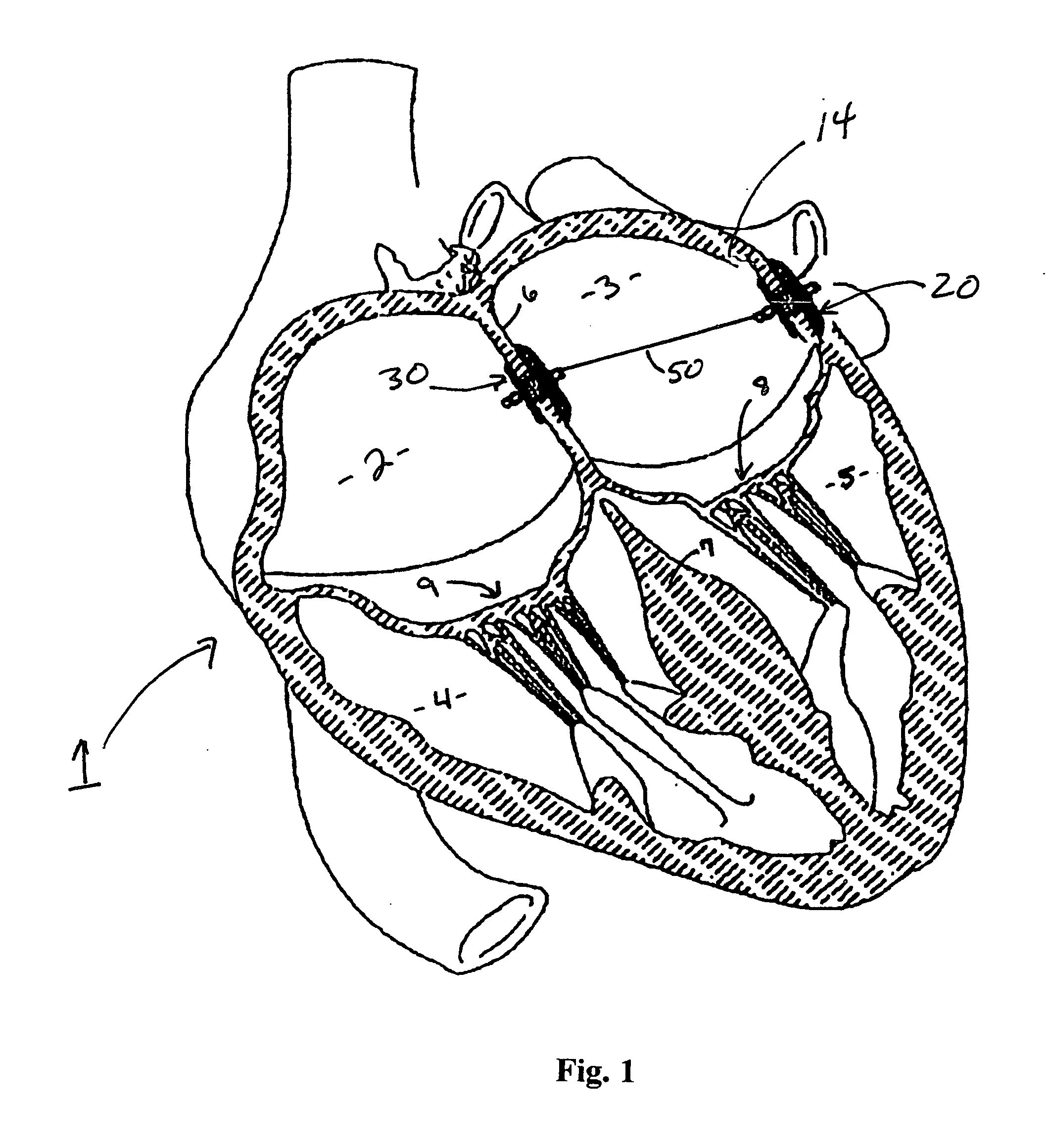
[0066] The human heart 1 includes four chambers, the right atrium 2, the left atrium 3, the right ventricle 4 and the left ventricle 5. The atrial septum 6 separates the right and left atria. The ventricular septum 7 separates the ventricles. The mitral valve 8 separates the left atrium 3 from the left ventricle 5. The tricuspid valve 9 separates the right atrium 2 from the right ventricle 4.

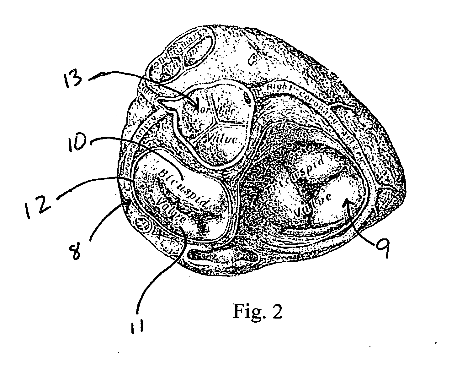
[0067] As shown in FIG. 2, the mitral valve 8, sometimes referred to as the bicuspid valve, is made up of two leaflets 10 and 11 partially surrounded by an annulus 12, a diaphanous incomplete ring around the valve. During left ventricular diastole, after pressure drops in the left ventricle 5 due to relaxation of the ventricular myocardium, the mitral valve 8 opens and blood travels from the left atrium 3 into the left ventricle 5. In a healthy heart, the mitral valve 8 closes completely and the aortic valve 13 opens so that blood is forced by contraction of the walls of the left ventricle 5 ou...


PUM


Abstract

A method and apparatus for providing safe and efficient transcatheter correction of the shape of heart chambers, valves, or other body members. The apparatus generally performs this correction by securing a tether to body surfaces. The attachment device includes an anchor comprising a wire braid and at least one clamp affixed to and constraining a portion of the wire braid, the clamp having threading for temporarily coupling the anchor to a delivery member and an internal lumen through which the tether can pass. The device also includes a locking member cooperating with the lumen to lock the tether to the clamp at a preselected position along the length of the tether.

Description

I. FIELD OF THE INVENTION [0001] The present invention relates to devices and methods for improving the function of a diseased heart. Such devices and methods are particularly useful for non-invasively treating structural conditions of the diseased heart that can lead to morbidity and early death. II. BACKGROUND OF THE INVENTION [0002] The human heart is a complex pumping system of contracting chambers and valves that causes blood to flow through the vascular system of the body. The healthy human heart beats on average more than 40 million times a year. Over time, disease and injury can cause damage to the heart. In a diseased heart, the chambers can become swollen and distended leading to cardiac inefficiency and heart failure. Such swelling can lead to damage to the electrical conduction paths in the heart that control its rhythm. Likewise, the annulus of the mitral valve of the heart can become distended such that the leaflets do not fully close permitting blood to regurgitate (i...

Claims


Application Information

Patent Timeline
IPC IPC(8): A61B17/08
CPCA61B17/00234A61B17/0401A61B17/0469A61B2017/00243A61B2017/00575A61F2/2487A61B2017/00606A61B2017/00619A61B2017/00623A61B2017/00783A61B2017/048A61B2017/00592
Inventor NELSON, DALEGU, XIAOPINGANDERSON, KENT
Owner AGA MEDICAL CORP MS US
Learn More