
Fractionator and Method of Fractionation

a technology of fractionator and fractionation method, which is applied in the field of fractionator and method of fractionation, can solve the problems of complex operation of pretreatment, difficulty in using ms for daily clinical investigations, and inability to carry out proteome analysis in clinical field, so as to facilitate detection of high efficiency, and low molecular weight proteins

Inactive Publication Date: 2007-10-18
TORAY IND INC
View PDF12 Cites 8 Cited by
  • Summary
  • Abstract
  • Description
  • Claims
  • Application Information

AI Technical Summary

Benefits of technology

[0039] Owing to the employment of the closed circuit apparatus, the fractionation device disclosed as the first invention can simply and efficiently carry out fractionation of high molecular weight proteins such as albumin from a raw liquid, particularly from a body fluid such as serum in a short time while preventing contamination of the analysis sample (recovered liquid of the fractionation device) and biohazard. Further, in the device of the invention, a portion of the device is disposed in a cartridge, the fractionation process of a next sample can easily be started.
[0040] According to the second invention, proteins with high molecular weights are efficiently removed from a solution containing a plurality of proteins with different molecular weights and a solution enriched with a trace of low molecular weight proteins can be obtained to make it possible to easily detect these low molecular weight proteins by mass spectrometry.
[0041] According to the third invention, proteins with high molecular weights are efficiently removed from a solution containing a plurality of proteins with different molecular weights and a trace of low molecular weight proteins can be recovered at a high efficiency.

Problems solved by technology

However, although the techniques and appliances have been improved swiftly, the present situation is not yet ready to simply and quickly carry out proteome analysis in a clinical field.
It is needed to fractionate and refine proteins of a clinical sample as pretreatment of mass analysis and the treatment still takes several days and the operation of the pretreatment is complicated and requires experiences and skills and that becomes a high obstacle against the clinical application.
Therefore, it must be said that at the present moment, MS is insufficient in practical applications for the purpose of obtaining analysis results within a time as short as possible for diagnosis and medical care in medical treatment fields and it becomes a significant cause of difficulty of utilization of MS for the daily clinical investigations.
However, they all have problems such as insufficiency of the separation and fractionation capability, unsuitability for a very small amount of a sample, contamination of chemical agents to be obstacles for mass spectrometry, and scanty in reproducibility.
However, they are complicated and time-consuming techniques and therefore, devices convenient and having high separation efficiency within a short time have been required.
In these years, a method of using a gel, Affi-Gel Blue, (reference to Non-Patent Document No. 4) and a method of using “Gradiflow” system (reference to Non-Patent Document No. 5) are reported as effective and improved albumin removal methods, however no technique further simplified and highly efficient for separation has not been reported yet.
Also, it cannot be denied that the Blue gel removes particularly only proteins with high molecular weights like albumin and at the same time also proteins to be subjected to proteome analysis.
As a filtration apparatus in which a liquid can be circulated, a filtration apparatus comprising a housing packed with a spirally rolled plane membrane is disclosed (reference to Patent Document 2), however the separation capability of the apparatus as it is not sufficient.
So far, no apparatus for which a countermeasure against the foreign substance contamination is performed has been made available.
Patent Document 3 discloses only a method but not a practical apparatus having a structure indispensable for protein separation.
In the case a plurality of different samples are treated, washing work is required to avoid contamination between respective analysis processes and consequently, it takes a time and in case, pathogens are contained in the samples, the pathogen leakage may occur during the treatment and it may possibly infect a worker with the pathogens.

Method used


Image

  • Fractionator and Method of Fractionation
  • Fractionator and Method of Fractionation
  • Fractionator and Method of Fractionation

Examples


examples

[0169] At first, Examples of the first invention will be described.

example a

First Invention

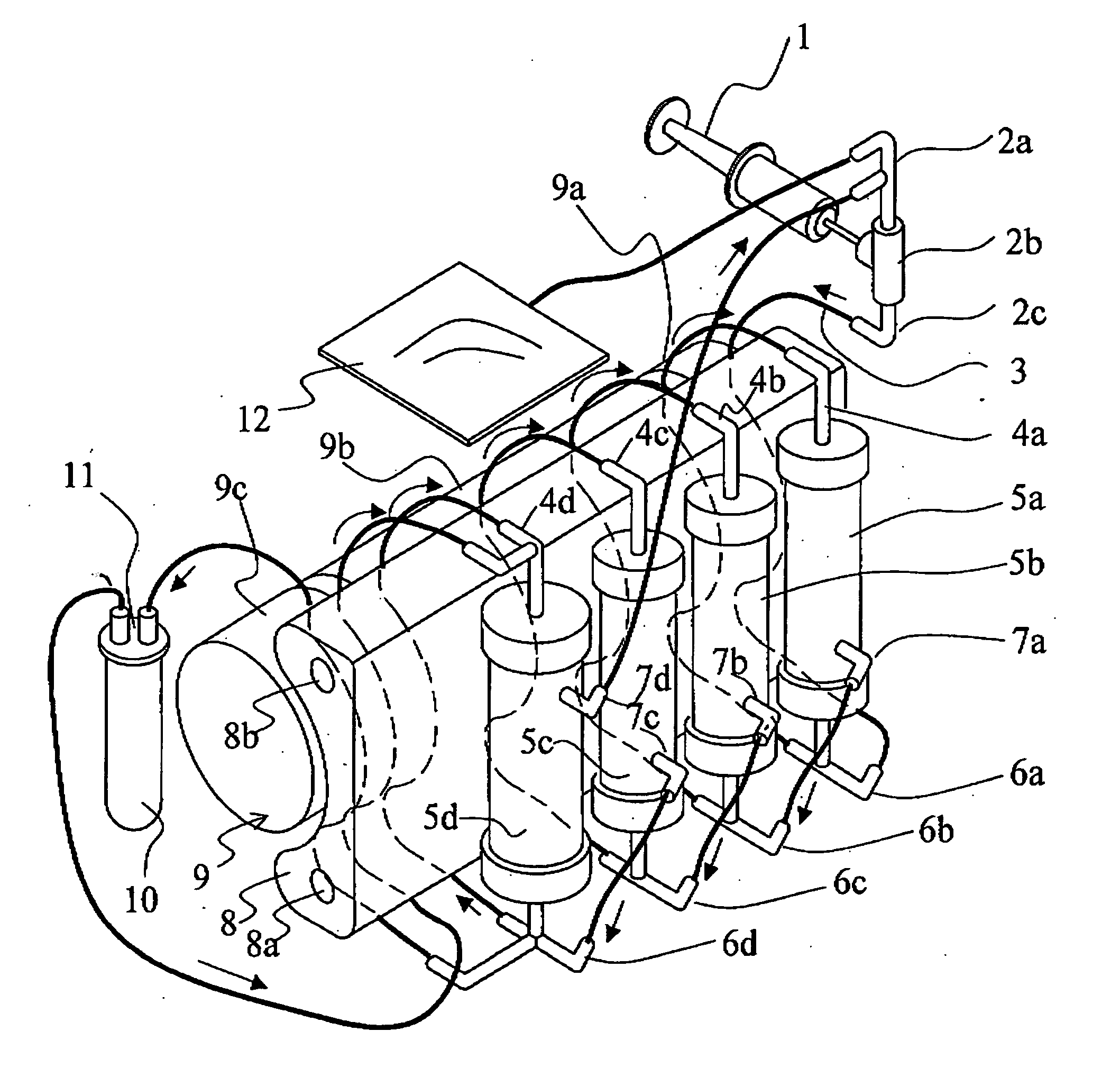
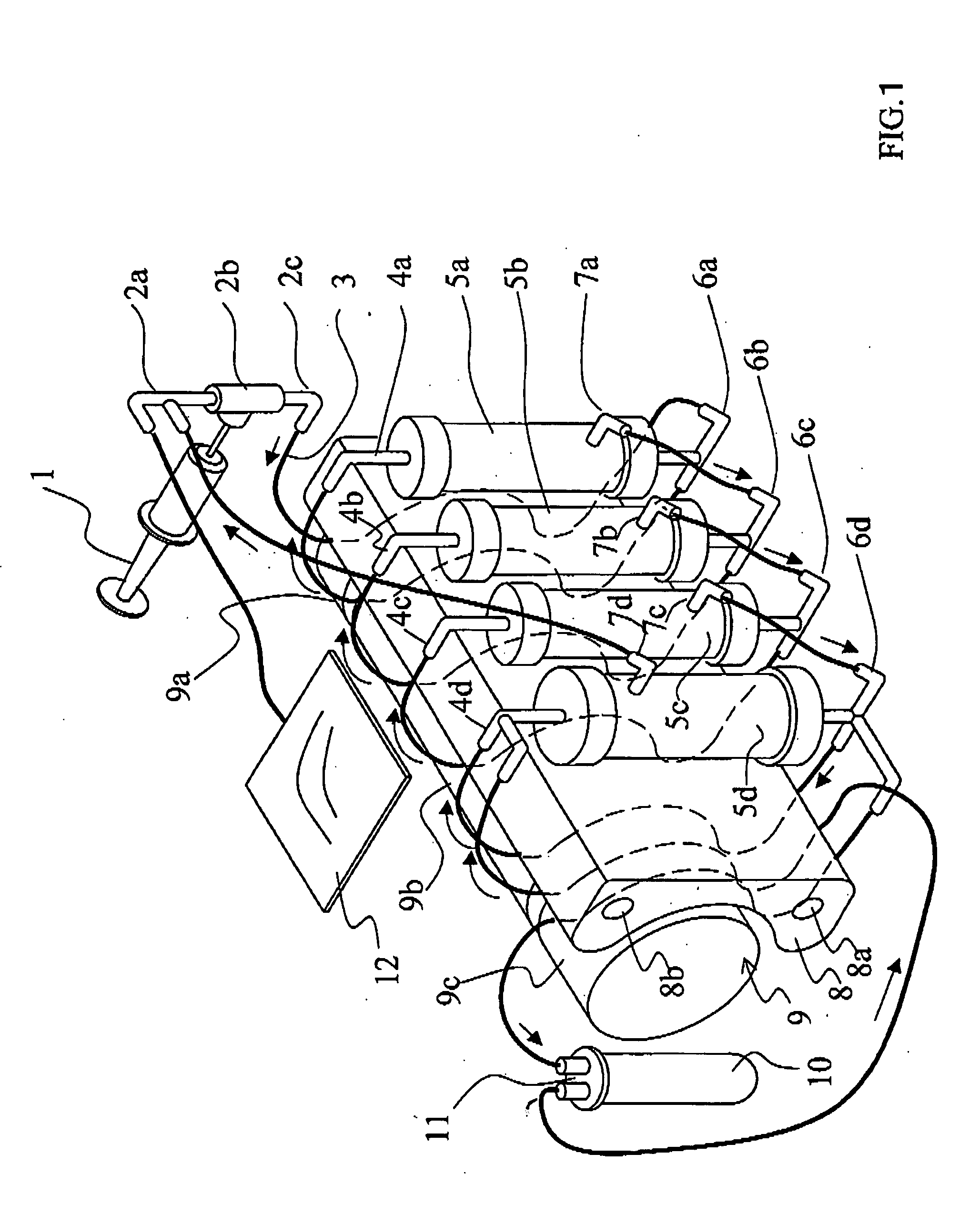
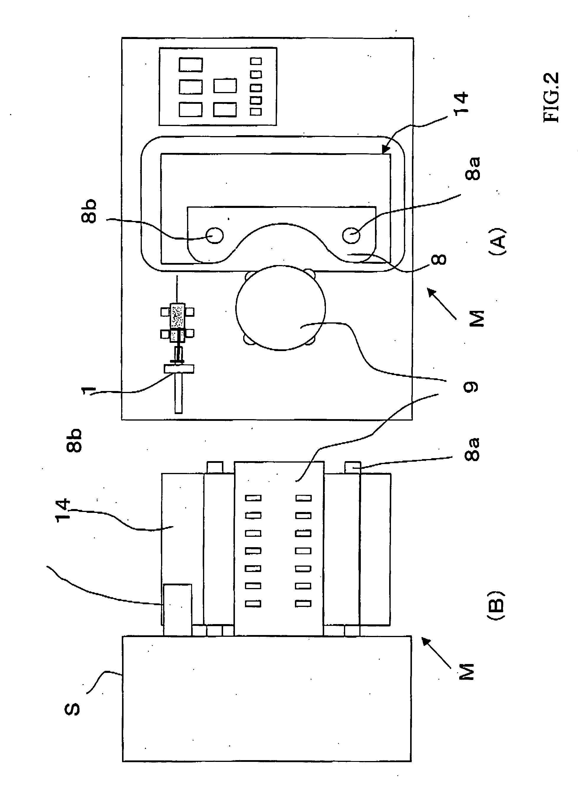
[0170]FIGS. 1 and 2 are explanatory drawing of a fractionation device of the invention. FIG. 1 shows the separation part is composed of three modules.

[0171] In FIG. 1, a three-way joint 2a and a joint 2c are connected to the rubber button 2b corresponding to the supply part. A flexible tube 3 connects the joint 2c and a lower nozzle 6a of hollow fiber membrane module 5a of the filtration part along the curved face of a multi-channel type squeezing member 8. Further a tube-equipped bag 12 is connected to the three-way joint 2a. Flexible tubes are connected to respective upper nozzles 4a, 4b, 4c, and 4d installed on the squeezing member, the filtration part hollow fiber membrane modules 5a, 5b, and 5c and a concentration part 5d. These tubes are laid along the curved face of the multi-channel type squeezing member 8 and respectively connected with the lower nozzles 6a, 6b, 6c, and 6d. Tubes are connected between a trunk lower nozzle 7a of the separation part hollow fi...

example 1

[0178] A hundred polysulfone hollow fibers were bundled and both ends were fixed in a glass tube type module case with an epoxy type potting agent in a manner the hollow parts of the hollow fibers were not closed to produce a mini-module. The mini-module had an inner diameter of about 7 mm and a length of about 17 cm and two dialysis ports similarly to a common hollow fiber membrane type dialyzer. The hollow fibers of the mini-module and the inside of the module were washed with distilled water.

[0179] After that, an aqueous PBS (Dulbecco PBS (−) manufactured by NISSUI PHARMACEUTICAL CO., LTD) solution was packed to obtain a hollow fiber membrane mini-module (hereinafter, referred to as mini-module 1 for short). After precipitates were removed from human serum (H1388, Lot 28H8550, manufactured by SIGMA) by centrifugation at 3000 rpm for 15 min, the resulting human serum was filtered with a 0.45 μm filter. One of the dialyzed liquid side of the mini-module 1 was caped and the other w...


PUM

PropertyMeasurementUnit
Temperatureaaaaaaaaaa
Fractionaaaaaaaaaa
Fractionaaaaaaaaaa

Abstract

The following is disclosed: (1) a membrane fractionator including a filtration section, a concentrating section, a recovery section and a liquid feed pump, wherein a flow channel connecting the filtration section, concentrating section and recovery section to each other constitutes a closed circuitry; (2) a method of biocomponent separation, characterized in that a sample derived from biocomponents is fed into an antibody adsorption membrane separation system having an antibody capable of adsorbing specified protein internally accommodated in the middle or a rear part of a membrane separation system that in the absence of antibodies adsorbing proteins, exhibits a permeation ratio between human alpha1-microglobulin and human albumin of 1.5 to 1000, thereby separating part of the biocomponents; and (3) a method of protein fractionation, comprising bringing a solution containing two or more types of proteins and water into contact with a hollow yarn separation membrane to thereby attain protein fractionation, characterized in that the fractionation solution contains an organic solvent.

Description

FIELD OF THE ART [0001] The invention relates to a method and a device for obtaining a sample with a changed composition from a biological component-containing solution, particularly from a raw liquid such as human blood, plasma, urine, or the like by fractionating biological molecules such as protein of the solution, particularly the raw liquid. Specifically, aiming to make clinical proteome analysis possible, the invention relates to a fractionation method and a fractionation device for obtaining a solution with a changed composition of biological components by removing components inhibiting detection of trace components, particularly high molecular weight proteins. PRIOR ART [0002] Recently, proteome analysis research proteomics has begun to draw attention as postgenome research. Since it is a very likely supposition that proteins, gene products, are more directly linked with symptoms of diseases than gene, it has been highly expected that research findings and achievements of pr...

Claims


Application Information

Patent Timeline
IPC IPC(8): C07K1/34B01D37/00B01D63/00B01D61/14B01D63/02B01D63/04C07K1/22
CPCB01D61/142B01D61/20B01D63/04B01D63/08C07K1/36B01D2319/022C07K1/22C07K1/34B01D2313/243
Inventor TANAHASHI, KAZUHIROKUMO, ICHIROKUROKI, NOBUYUKISUGAYA, HIROYUKIYAMADA, SATOKOWADA, SHIGEHISAJUNG, GIMANKURODA, TOSHIHIKOSEKIGUCHI, SHUJI
Owner TORAY IND INC
Learn More