
Device and Method for Administering Therapeutic Agents

a technology of therapeutic agents and devices, applied in the field of devices and methods for administering therapeutic agents, can solve the problems of not being able to achieve satisfactory results of these methods, not being able to prevent cauti with the cuff, and not being able to achieve the above mentioned methods, so as to achieve easy control of time, place and concentration, and low systemic toxicity. , the effect of simple and safe solution

Inactive Publication Date: 2007-10-11
NITRICARE
View PDF20 Cites 69 Cited by
  • Summary
  • Abstract
  • Description
  • Claims
  • Application Information

AI Technical Summary

Benefits of technology

[0104] Yet another advantage of the present invention is the in situ production of the active substance, the LMAC, when the at least one component and the second component are combined in the cuff. There is no loss of antibacterial activity during transportation or delivery.
[0105] A particular advantage lies in the fact that the inventive device and method can be easily introduced in clinical practice.
[0106] The present invention will now be further described in the following non-limiting example. EXAMPLES

Problems solved by technology

Systemic administration of drugs often causes side effects on organs and tissue not intended for treatment.
However, those coated catheters mainly affect the infections caused by Gram positive organisms or yeasts adhering to the surface of the catheter.
None of these methods have given satisfactory results and new better ways to inhibit nosocomial infections would be valuable to replace or complement existing methods.
None of the above mentioned methods for preventing CAUTI relate to the cuff and preventing biofilm formation and bacterial growth on the surface of the cuff.
For example, in silver coated catheters the inflatable cuff may be difficult to coat and may remain a particularly susceptible site for infection.
The fact that the release of the antibiotics is driven by osmosis renders the antibacterial effect difficult to control.
The water content of the urine is highly variable depending on the present fluid balance of the body, which can lead to an uncontrolled and uneven distribution of the antibiotics.
Furthermore, the release of the antibiotic to the outside of the bladder will be a slow process, exposing the bacteria to the antibiotic during a prolonged time.
This increases the risk of development of antibiotic resistant bacteria.

Method used

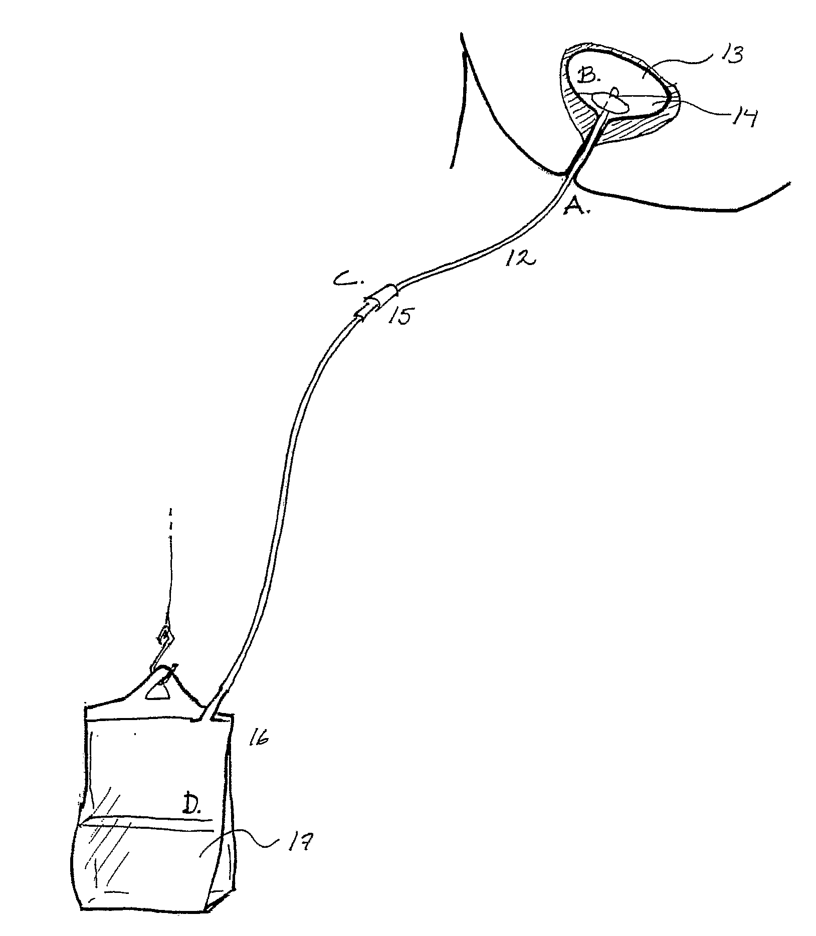
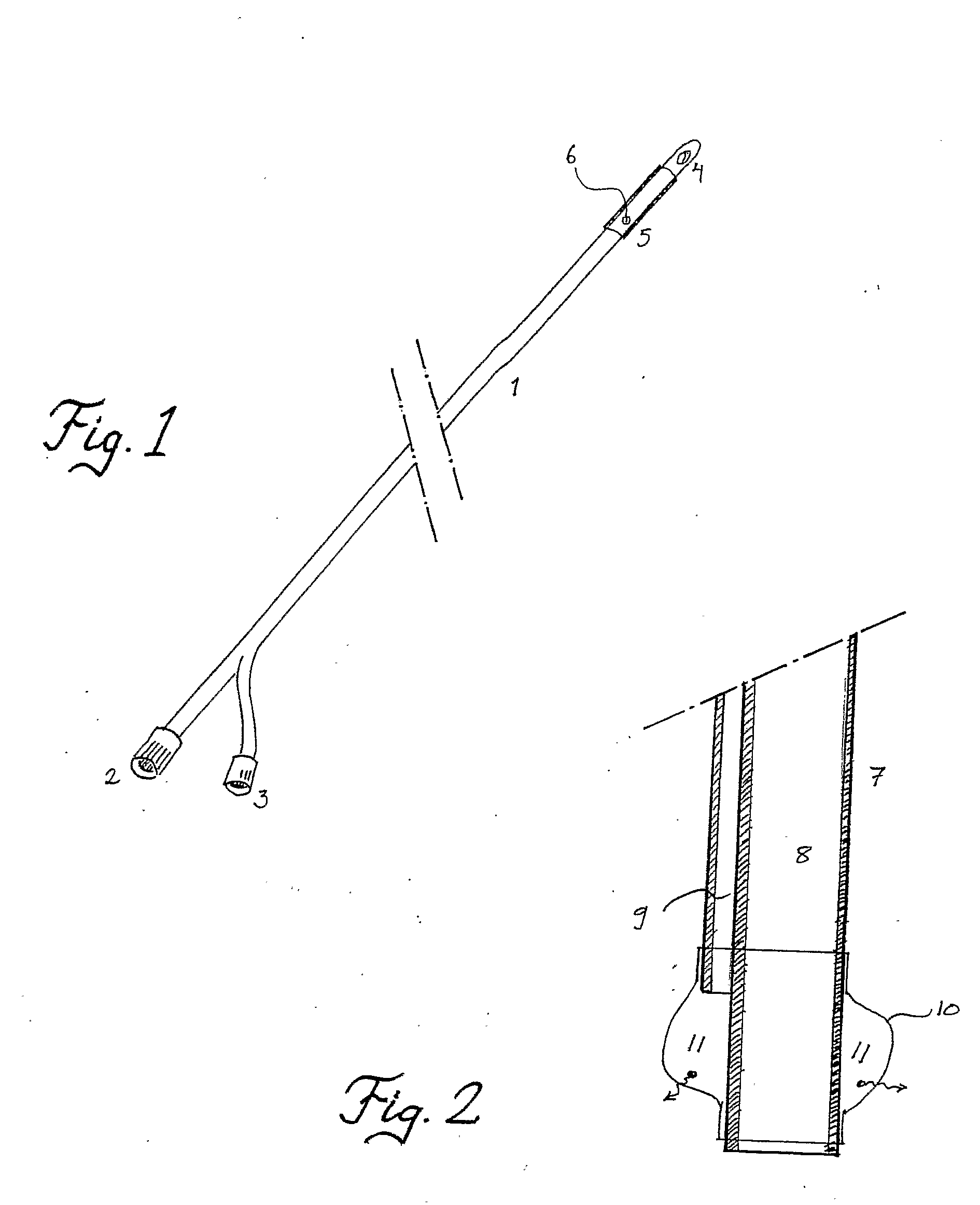
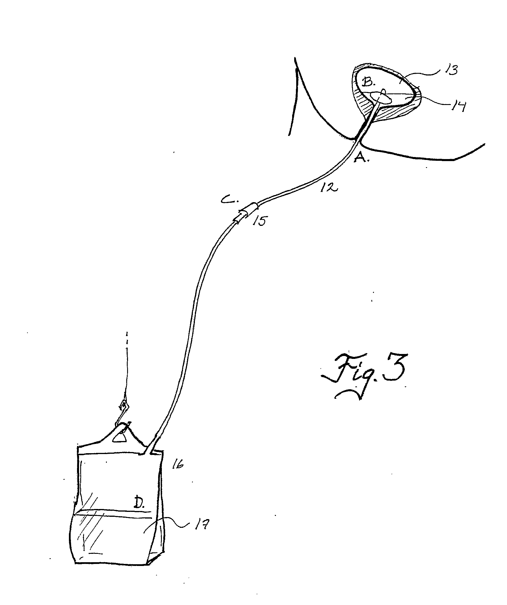

Image

  • Device and Method for Administering Therapeutic Agents
  • Device and Method for Administering Therapeutic Agents
  • Device and Method for Administering Therapeutic Agents

Examples


example 1

Incubation of an E. coli Strain in a Solution of Ascorbate and Sodium Nitrate

1.1 Materials & Methods

[0107] An E. coli strain isolated from urine of a patient with lower urinary tract infection was used. A 50 ml glass bottle with a narrow neck (FIG. 4) was filled with fresh urine (pH 6.5) from a healthy volunteer. The urine was inoculated with the E. coli strain to a final density of 105 colony forming units / ml. Then an all-silicone urinary catheter was inserted in the bottle and the cuff was filled with a solution comprising: [0108] 1. Saline+ascorbate (20 mM)+HCl to a final pH of 2.0 (control; n=3) or [0109] 2. Saline+ascorbate (20 mM)+HCl+sodium nitrite (2 mM), pH 2 (nitrite; n=3)

[0110] The expanded cuff was fixed at the neck of the bottle to prevent leakage of urine when the bottle was turned up side down. The glass bottle was then incubated in 37° C. for 10 h after which growth of E. coli in the surrounding urine was monitored by optical density (OD) at 540 nm on a Spectrama...

example 2

Viable Counts

2.1 Materials & Methods

[0113] An E. coli isolated from a patient with urinary tract infection and a reference strain, E. coli ATCC 25992 were used in this study. An overnight culture was added to 25 ml of urine to a final density of 105 CFU / ml. The urine was placed in long-necked 50 ml flasks with a shape resembling the urinary bladder and the urethra. An all-silicone catheter (Argyle, Sherwood Medical, Tullamore, Ireland) was placed in the flask and the retention balloon was filled with 10 ml of saline containing ascorbic acid (10 mM) and sodium nitrite (5 mM). The pH of the solution was adjusted to 2.5 using hydrochloric acid (3 M). Ascorbic acid and nitrite were prepared and mixed immediately before administration. Ascorbic acid solution alone (pH 2.5) was used as control. After filling the retention balloons the catheter was gently pulled outwards and fixed at the neck whereby the flask opening was sealed off. Then the flasks were turned up side down and incubate...

example 3

Drug Release Kinetics

3.1 Materials & Methods

[0117] The kinetics of NO release from the retention balloon was monitored in separate experiments. The catheter was placed in the flask and after filling the retention balloon with the ascorbic acid / nitrite solution the flask was closed. Synthetic NO-free air was flushed via an inlet at a rate of 4.5 L / min and headspace NO was continuously measured from an outlet by a rapid-response chemiluminescence system (Aerocrine AB, Stockholm, Sweden).

3.2 Results

[0118] The NO release rate from the balloons peaked initially and then decreased with a half-time of about 30 min, see FIG. 8. The results prove the concept underlying the invention and show the utility of the device and method for the treatment of urinary tract infections.


PUM


Abstract

The present invention discloses a device and a method for reducing the risk of hospital-acquired infections, nocosomial infections, originating from the insertion of the device and / or use of the device in the body of a human or animal. The invention in particular relates to a device releasing at least one low molecular antimicrobial compound permeating the device thereby exerting its antimicrobial effect also on the outside of the device.

Description

[0001] The present invention concerns a device and method for administering therapeutic agents, e.g. for the treatment or prevention of a disease, such as but not limited to infectious diseases, inflammatory diseases, cancer etc., or for preventing or reducing the incidence of nosocomial infections in humans and animals having an invasive medical device inserted into the body, in particular catheter-associated urinary tract infections. More specifically, the invention relates to a device having means for releasing low molecular therapeutic agents, such as anticancer drugs, anti-inflammatory, antiviral or antimicrobial compounds that permeate to the adjacent tissue and / or body cavity. BACKGROUND [0002] In the field of medicine, a distinction can be made between local and systemic treatments, and between invasive and non-invasive interventions, in the prevention, treatment or alleviation of an abnormal function or state involving any structure, part or system of a living organism. Sys...

Claims


Application Information

Patent Timeline
IPC IPC(8): A61M29/00A61F2/958A61J15/00A61M31/00
CPCA61J15/0015A61J15/0042A61M31/00A61M25/10A61P29/00A61P31/00A61P31/02A61P31/04A61P35/00A61P41/00A61P43/00A61M29/02
Inventor LUNDBERG, JONWEITZBERG, EDDIE
Owner NITRICARE
Learn More