Differentiation of human embryonic stem cells to cardiomyocytes

a technology cardiomyocytes, applied in the field of human embryonic stem cells, can solve the problems of wide spread of cell death and loss of function

Inactive Publication Date: 2007-07-12
MUMMERY CHRISTINE LINDSAY +2
View PDF0 Cites 31 Cited by
  • Summary
  • Abstract
  • Description
  • Claims
  • Application Information

AI Technical Summary

Benefits of technology

[0009] The stem cells suitable for use in the present methods may be derived from a patient's own tissue. This would enhance compatibility of differentiated tissue grafts derived from the stem cells with the patient. In this context it should be noted that hES cells can include adult stem cells derived from a person's own tissue. Human stem cells may be genetically modified prior to use through introduction of genes that may control their state of differentiation prior to, during or after their exposure to the embryonic cell or extracellular medium from an embryonic cell. They may be genetically modified through introduction of vectors expressing a selectable marker under the control of a stem cell specific promoter such as Oct-4. The stem cells may be genetically modified at any stage with a marker so that the marker is carried through to any stage of cultivation. The marker may be used to purify the differentiated or undifferentiated stem cell populations at any stage of cultivation.
[0037] Vital fluorescent staining with ryanodine or antibodies against cell surface α1c ion channels allowed differentiated cardiomyocytes of the invention to be identified in mixed cultures. This may provide a means of isolating cardiomyocytes for transplantation without genetic manipulation or compromising their viability.
[0049] Conditions for obtaining differentiated embryonic stem cells are typically those which are non-permissive for stem cell renewal, but do not kill stem cells or drive them to differentiate exclusively into extraembryonic lineages. A gradual withdrawal from optimal conditions for stem cell growth favours differentiation of the stem cell to specific cell types. Suitable culture conditions may include the addition of DMSO, retinoic acid, FGFs or BMPs in co-culture which could increase differentiation rate and / or efficiency.

Problems solved by technology

Oxygen deprivation and subsequent reperfusion initiates irreversible cell damage, eventually leading to widespread cell death and loss of function.

Method used


Image

  • Differentiation of human embryonic stem cells to cardiomyocytes
  • Differentiation of human embryonic stem cells to cardiomyocytes
  • Differentiation of human embryonic stem cells to cardiomyocytes

Examples


Embodiment Construction

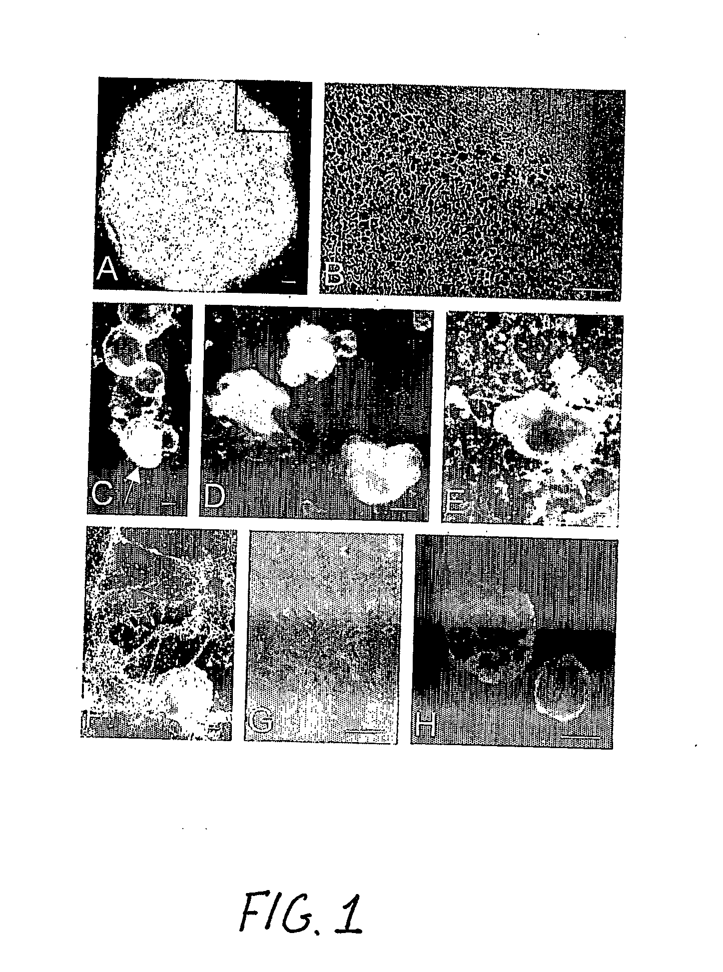
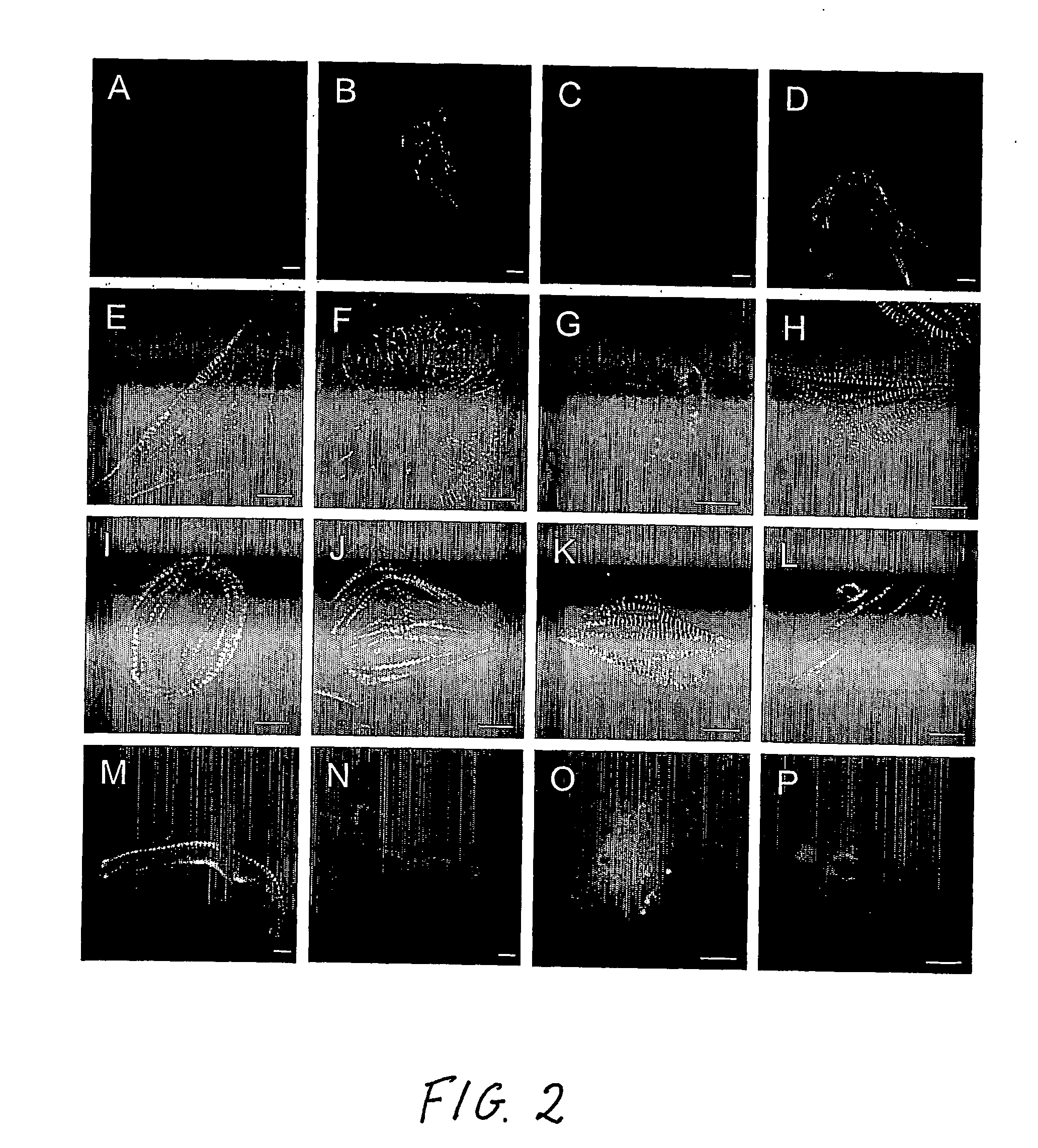
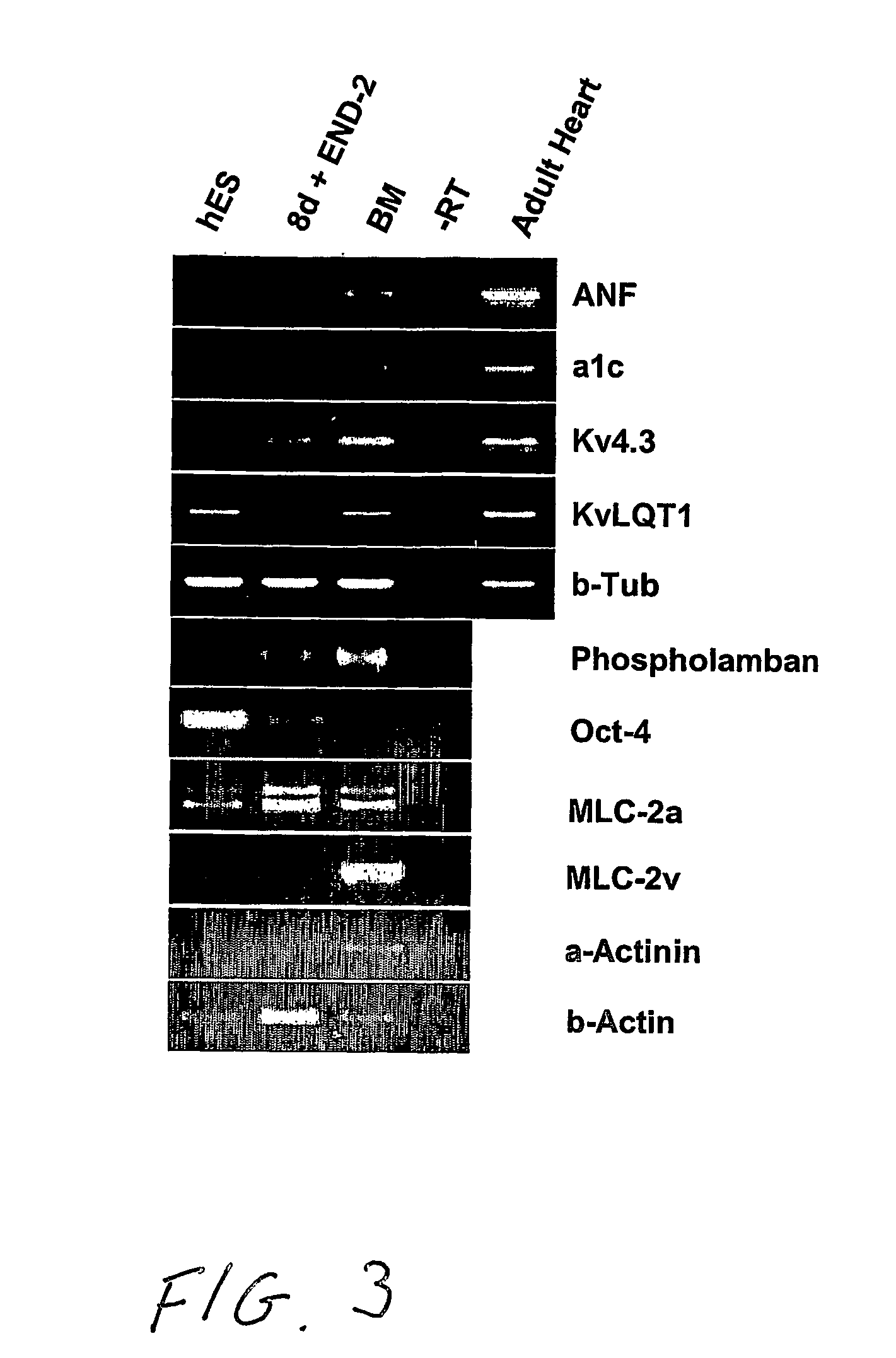
[0006] Human embryonic stem cells co-cultured with mouse visceral endoderm (VE)-like cells form beating muscle cells, expressing cardiac specific sarcomeric proteins and ion channels. Direct comparison of electrophysiological responses demonstrated that the majority resembled human fetal ventricular cells in culture; a minority had an atrial phenotype. Both fetal and hES-derived cardiomyocytes in culture were functionally coupled through gap junctions. This is the first demonstration of induction of cardiomyocyte differentiation in hES cells that do not undergo cardiogenesis spontaneously.

[0007] Here, we demonstrate that co-culture of pluripotent hES cell lines with END-2 cells induces extensive differentiation to two distinctive cell types from different lineages. One is epithelial and forms large cystic structures staining positively for alpha-fetoprotein and is presumably extraembryonic visceral endoderm; the others are grouped in areas of high local density and beat spontaneous...


PUM


Abstract

A method for inducing cardiomyocyte differentiation of a hES cell, the method comprising co-culturing the hES cell with a cell excreting at least one cardiomyocyte differentiation inducing factor or with an extracellular medium therefrom, under conditions that induce differentiation, cells and cell populations so produced, and uses of the cells.

Description

TECHNICAL FIELD [0001] The present invention relates to human embryonic stem cells (hES) and their differentiation. hES cells can give rise to cardiomyocytes that are a better model for studying human physiology than murine embryonic stem (mES) cell-derived cardiomyocytes or primary cardiomyocytes from adult or fetal mice. hES cells can give rise to cardiomyocytes which provide a model of relevance to the study of human cardiac disease. hES cells can give rise to normal and mutant cardiomyocytes suitable for testing drugs. BACKGROUND ART [0002] Cardiomyocytes have potential in restoring heart function after myocardial infarction or in heart failure. Human embryonic stem (hES) cells are a potential source of transplantable cardiomyocytes but detailed comparison of hES derived cardiomyocytes with primary human cardiomyocytes is necessary before transplantation into patients becomes feasible. [0003] Ischemic heart disease is the leading cause of mortality in the western world. Oxygen d...

Claims


Application Information

Patent Timeline
IPC IPC(8): C12N5/08C12N5/077
CPCC12N5/0657C12N2506/02C12N2502/02A61P9/00
Inventor MUMMERY, CHRISTINE LINDSAYDOEVENDANS, PETRUS ADRIANUS FREDERIK MARIATERTOOLEN, LEON GERARDUS JOSEPH
Owner MUMMERY CHRISTINE LINDSAY
Why Eureka
Try Eureka