
Binding layer and method for its preparation and uses thereof

a technology applied in the field of binding layer and matrices, can solve the problem that the binding was practically insufficient for the performance of an interaction assay

Active Publication Date: 2007-05-24
BIO RAD HAIFA
View PDF7 Cites 13 Cited by
  • Summary
  • Abstract
  • Description
  • Claims
  • Application Information

AI Technical Summary

Benefits of technology

[0014] According to the current invention, provided herein are improved binding layers, having significant benefits for use in bioassays and biosensors when compared with known and currently used matrices.
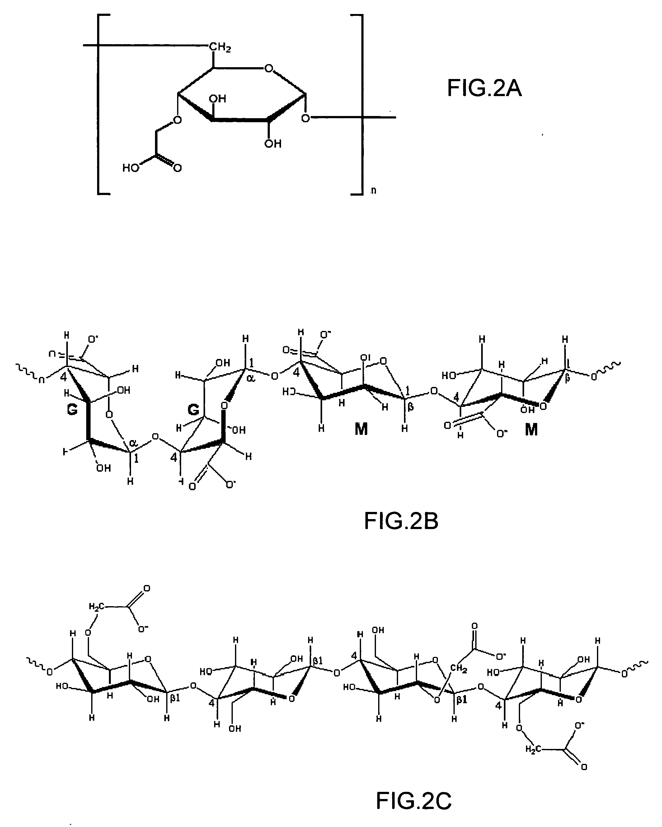
[0017] Consequently, these layers are capable of more efficient binding of ligand molecules. For example, the binding density of an antibody was doubled when CGs (as in prior-art layers) having an oxygen at the i-position (RO—group bound to the β-carbon), were transformed to easily-activated CGs.
[0018] More surprisingly, it was found that the improved layers can exhibit not only a more efficient ligand binding but also higher ligand activity, resulting in even more sensitive assays.
[0019] Furthermore, the electrostatic charge of the provided layers can be minimized efficiently upon subsequent processes of activation and deactivation. It will be demonstrated that a layer containing easily-activated CGs was neutralized using a process that showed almost no effect on similar layers containing the commonly used CGs, namely CGs which have oxygen groups substituted at their α-carbon.
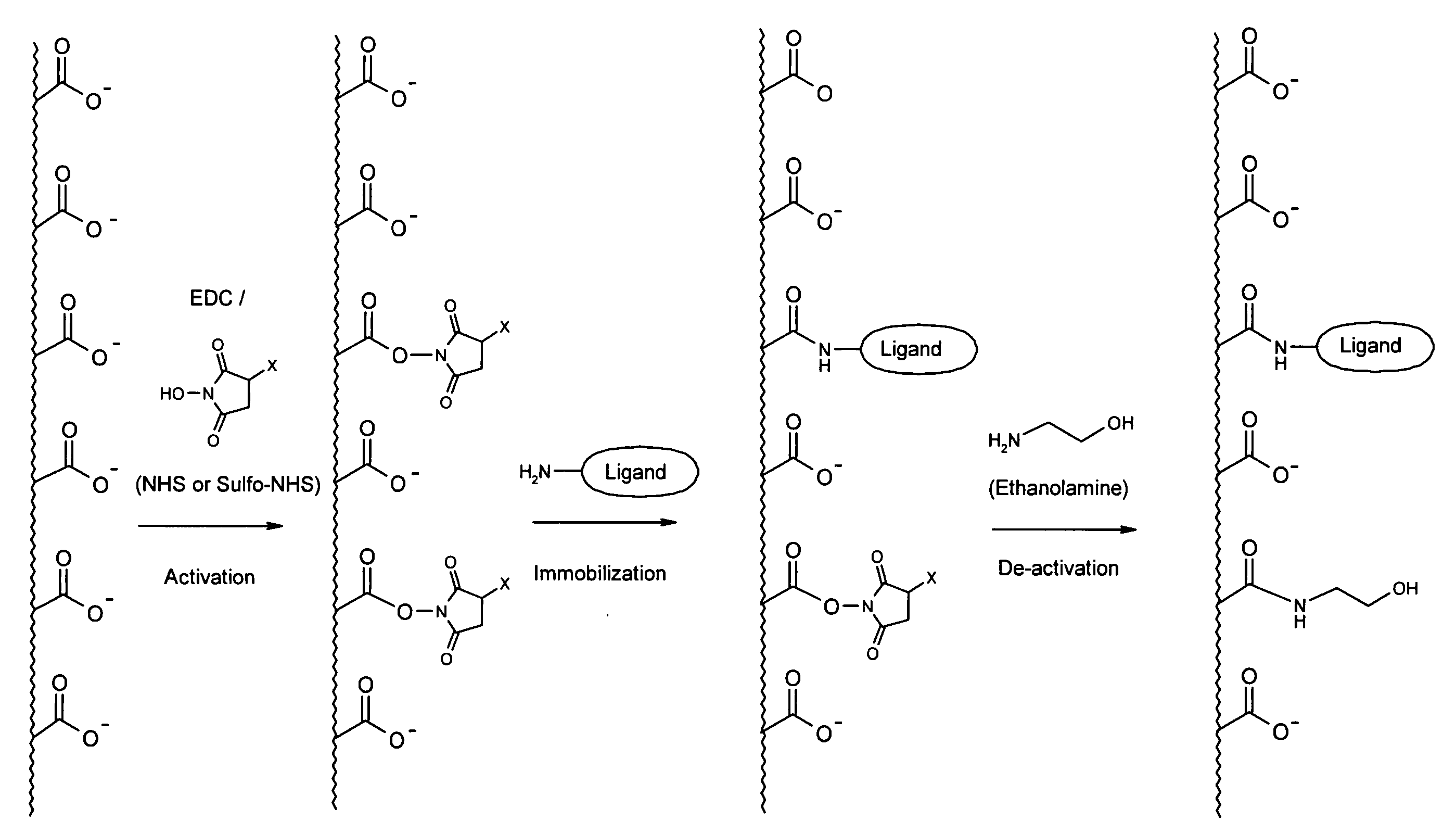
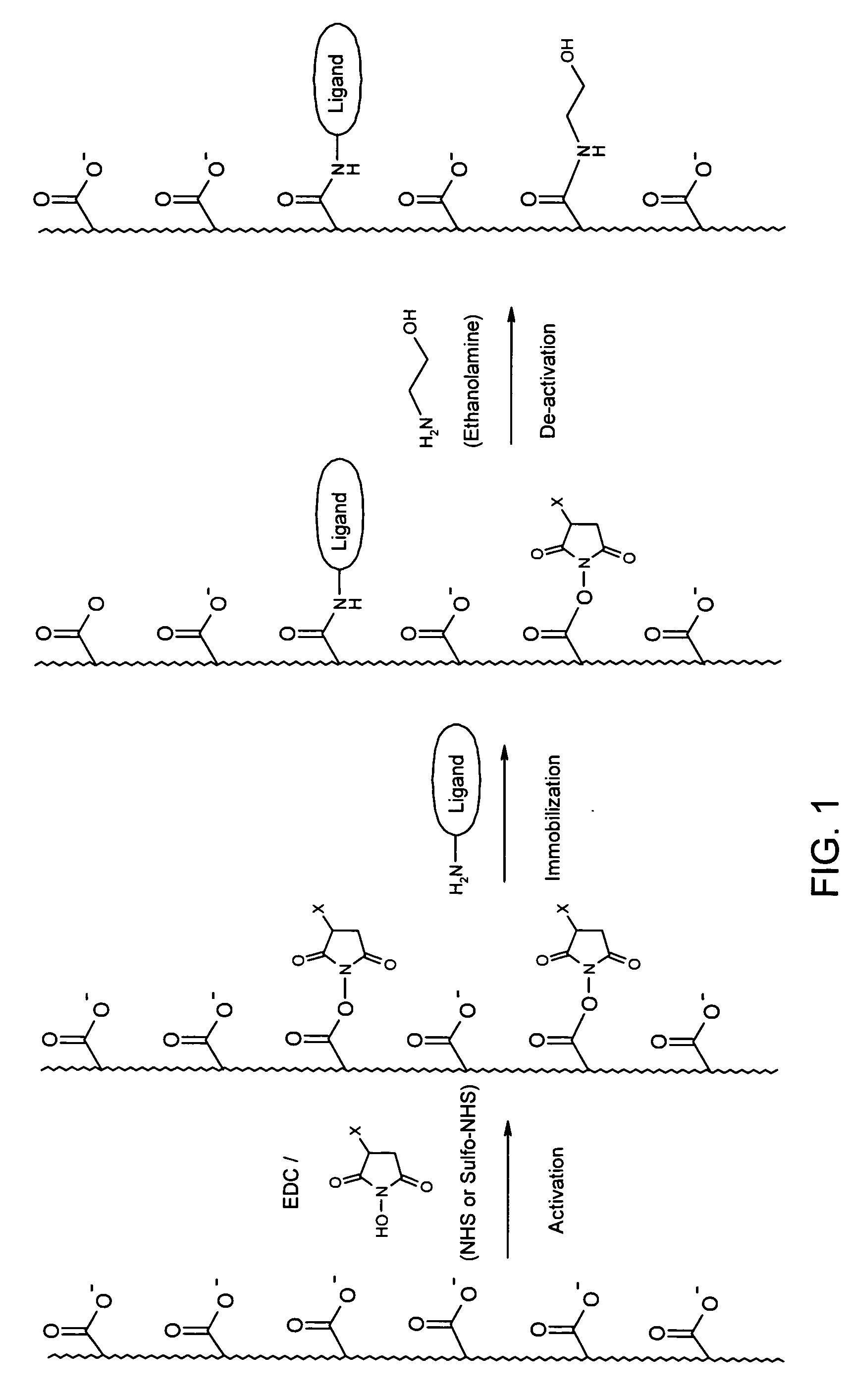
[0050] As will be demonstrated herein binding layers based on polysaccharides having easily-activated CGs have significant benefits over the commonly-used matrices having CGs with electron withdrawing group such as RO- bound to the α-carbon.

Problems solved by technology

In some cases, the binding was practically insufficient for performance of an interaction assay.
The remaining charge is undesired at the interaction assay stage, since charged analyte molecules might interact non-specifically with the layer, causing possible interruptions and distortions in the assay results, as familiar to any person skilled in the art.

Method used


Image

  • Binding layer and method for its preparation and uses thereof
  • Binding layer and method for its preparation and uses thereof
  • Binding layer and method for its preparation and uses thereof

Examples


example 1

Preparation of Binding Layers

[0072] Thin layers of CMD, alginic acid, carboxymethyl cellulose or poly(acrylic acid) were attached to the gold surface of ProteOn SPR sensor chips, using a technology adopted from Langmuir 2001, 17, 8336-8340.

[0073] Briefly, each of the polymers was dissolved in aqueous solution and reacted with cystamine dihydrochloride in the presence of EDC and NHS, under conditions in which a few percent of the CGs of the polymer were modified with the cytamine dimer. Then, the cystamine disulfide bonds were reduced by tetra(carboxyethyl phosphine) and the solution was purified by dialysis. The product was an aqueoussolution of the polymer, which now contained thiol end groups enabling its attachment to the gold surface.

[0074] The sensor chips were immersed in an aqueous solution containing the cystamine-modified polymers for 24 hours. The structure of the coating was varied by changing various parameters, such as the degree of cystamine modification, the concen...

example 2

Coupling Efficiency of a Representative Protein

[0078] Table 2 shows examples of ligand densities after adsorption or immobilization of Rabbit IgG antibody to five binding layers under similar conditions. The activation procedure included exposure to a solution of 0.2 M EDC and 0.05 M NHS or sulfo-NHS (7 min injection). The adsorption / immobilization of protein were done by exposure to a solution of 50 ug / ml Rabbit IgG in 10 mM sodium acetate buffer, pH 4.5 (6 min injection). Finally, the activated layers were deactivated by exposure to 1 M ethanolamine hydrochloride, pH 8.5 (5 min injection).

TABLE 2Ligand densities after adsorption or immobilization of Rabbit IgGantibody.AdsorptionBindingBindingcapacityafterafter EDC / LayerModifi-(noEDC / NHSsulfo-NHS#Polymercationactivation)activationactivation1Poly(acrylicNone6,000 RU5,100 RU6,000 RUacid)2Alginic acidNone6,100 RU3,000 RU3,100 RU3CarboxymethylNone5,900 RU2,900 RU3,000 RUcellulose4CarboxymethylNone6,000 RU3,000 RU3,100 RUdextran2EA...

example 3

Coupling Efficiency of Low-PI Protein

[0081] It is known that proteins with low isoelectric point (PI) are difficult to immobilize. Such proteins should be dissolved in a buffer with relatively low pH to render their positive charge and thus their electrostatic adsorption to the layer. Though, at low pH values, the negative charge of the CGs in the layer itself is decreased, and therefore the electrostatic attraction is weakened.

[0082] For example, it was reported that protein pepsin, which has a PI of 3.0, exhibited only negligible binding (70 RU) to CMD layer after standard EDC / NHS activation (Anal. Biochem. 1991, 198, 268-277). The results of a similar experiment using a binding layer based on alginic acid modified with β-alanine is shown in Table 2, layer 5E. These results show a much higher binding: 750 RU after EDC / NHS activation and 2050 RU upon EDC / sulfo-NHS activation. This advantage should be related to the use of easily-activated CGs, since the adsorption capacity of lay...


PUM


Abstract

The invention provides a binding layer comprising a polysaccharide substituted by carboxylic groups or derivatives thereof exhibiting high performance in the binding of ligand molecules and in the interaction thereof with analyte molecules. A method for the preparation of the binding layer and for the assaying of various analyte molecules is also provided.

Description

[0001] This application claims the benefit of prior U.S. provisional patent application No. 60 / 730,375 filed Oct. 27, 2005, the contents of which are hereby incorporated by reference in their entirety.FIELD OF THE INVENTION [0002] The present invention generally concerns binding layers and matrices for use in bioassays and biosensors. BACKGROUND OF THE INVENTION [0003] Biosensors have been developed to detect a variety of biomolecular complexes including oligonucleotides, antibody-antigen interactions, hormone-receptor interactions, and enzyme-substrate interactions. In general, biosensors consist of two components: a highly specific recognition element and a transducer that converts the molecular recognition event into a quantifiable signal. Such signal transduction may be accomplished by many methods, including fluorescence, interferometry, and gravimetry. [0004] Direct methods that do not require labeling of analytes with fluorescent compounds are of interest due to the relative ...

Claims


Application Information

Patent Timeline
IPC IPC(8): C12M1/34
CPCG01N33/548G01N33/54353
Inventor NIMRI, SHAI
Owner BIO RAD HAIFA
Learn More