Shotgun scanning
a scanning and shotgun technology, applied in the field of shotgun scanning, to achieve the effect of being ready to adap
- Summary
- Abstract
- Description
- Claims
- Application Information
AI Technical Summary
Benefits of technology
Problems solved by technology
Method used
Image
Examples
example 1
Shotgun Scanning
[0174] Experimental: A phagemid pW1205a was constructed using the method of Kunkel (Kunkel et al., 1987, Methods Enzymol. 154:367) and standard well known molecular biology techniques. Phagemid pW1205a was used as the template for library construction. pW1205a is a phagemid for the display of hGH on the surface of filamentous phage particles. In pW1205a, transcription of the hGH-P8 fusion is controlled by the IPTG-inducible Ptac promoter (Amman, E. and Brosius, J., 1985, Gene 40, 183-190). pW1205a is identical to a previously described phagemid designed to display hGH on the surface of M13 bacteriophage as a fusion to the amino terminus of the major coat protein (P8), except for the following changes. The mature P8 encoding DNA segment of pW1205a had the following DNA sequences for codons 11 through 20 (other residues fixed as wild-type):
TAT GAG GCT CTT GAG GAT ATT GCT ACT AAC (SEQ ID NO 1)
This segment encodes the following amino acid sequence:
YEALEDIATN (SEQ ...
example 2
[0207] A library was constructed using pW1205a as the template, exactly as described in Example 1, except that the following mutagenic oligonucleotides were used:
Oligo 1 (mutate hGH codons 41, 42, 45, and 48): 5′-ATC CCC AAG GAA CAG ARM TMC TCA TTC TYG CAG AAC YCT CAG ACC TCC CTC TGT TTC-3′ (SEQ ID NO 7)
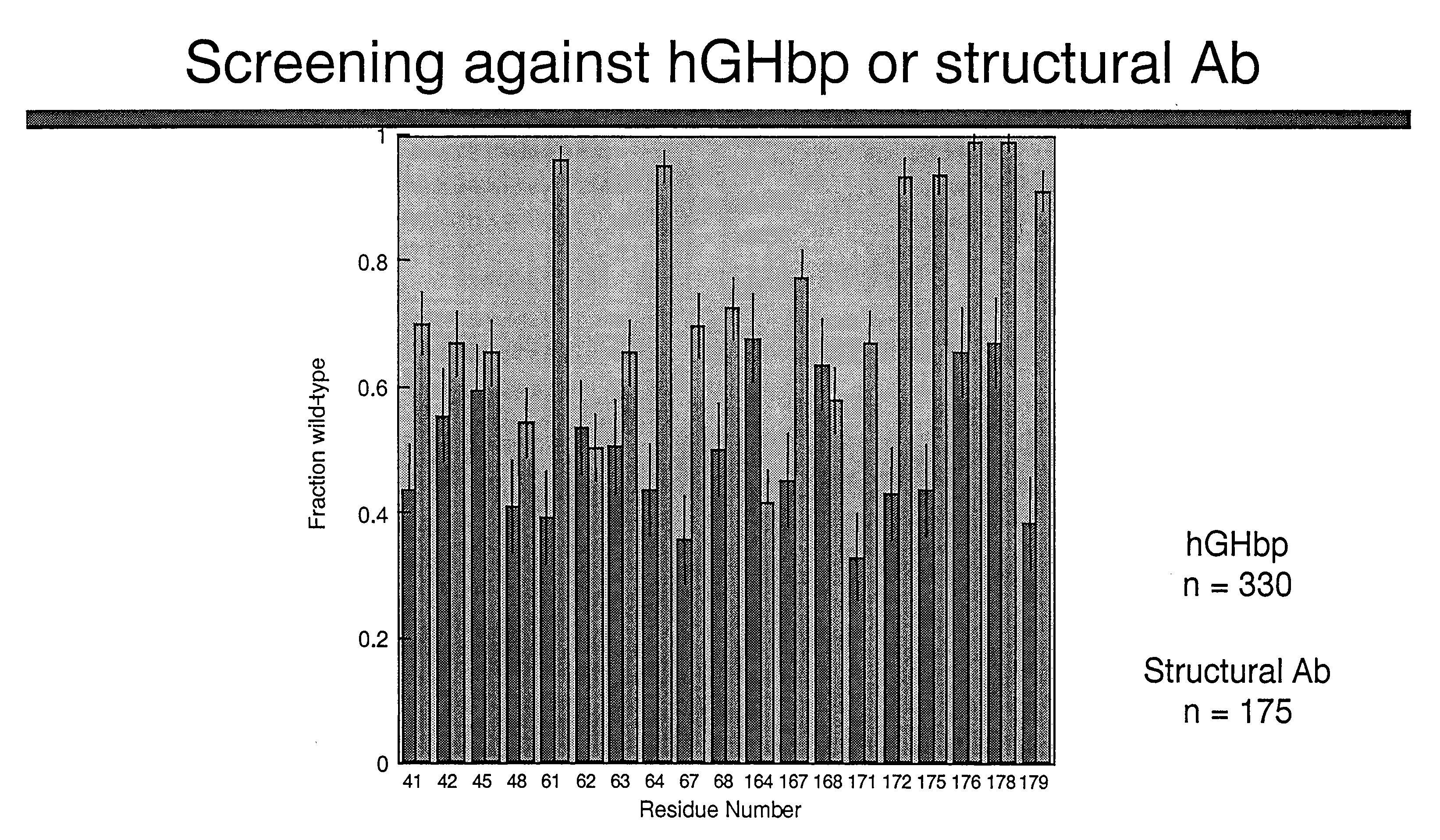
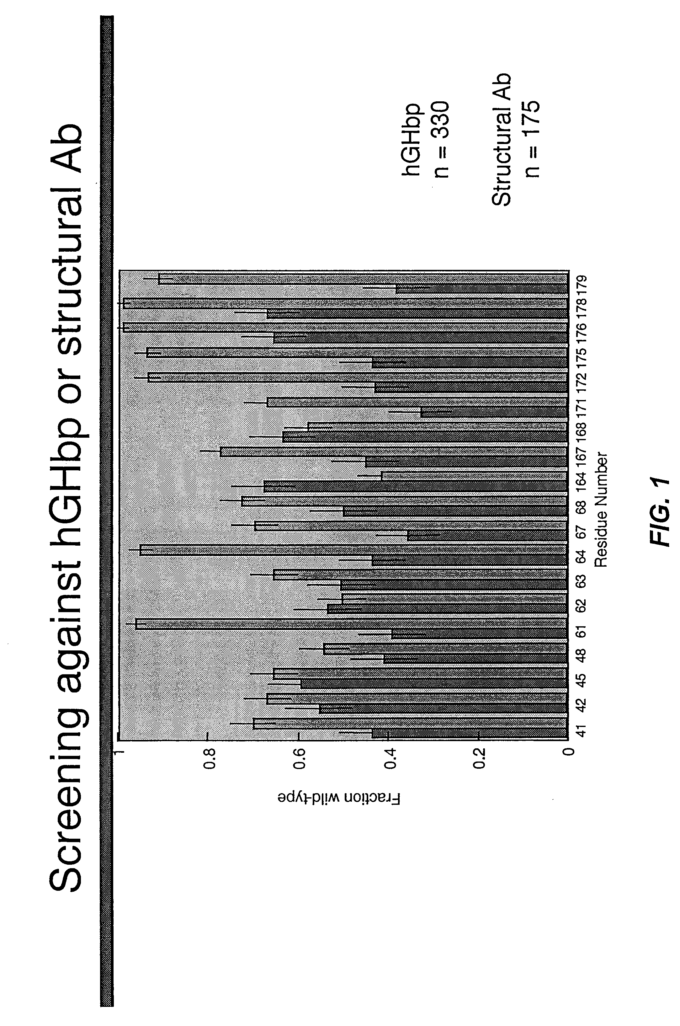
Oligo 2 (mutate hGH codons 61, 62, 63, 64, 67, 68): 5′-GAA TCG ATT CCG ACA YCT TCC ARC MGT GAG GAA WCG YMG CAG AAA TCC AAC CTA GAG-3′ (SEQ ID NO 8)
Oligo 3 (mutate hGH codons 164, 167, 168, 171, 172, 174, 175, 176, 178, 179): 5′-AAC TAC GGG CTG CTC TMC TGC TTC MGT ARM GAC ATG KMC ARM GTC KMG WCG TYC CTG MGT AKC GTG CAG TGC CGC TCT-3′ (SEQ ID NO 9)
[0208] The resulting library contained hGH variants in which the indicated codons were replaced by degenerate codons as described in Table 6. The library contained 2.1×10¹⁰ unique members. The library was sorted against either hGHbp or an anti-hGH antibody as described above and the resulting selectants were ...
example 3
Homolog Shotgun Scan of hGH
[0210] Standard molecular biology techniques were used to construct phagemid pW1269a. Phagemid pW1269a is identical to phagemid pW1205a (example 1) except that codons 14, 15, and 16 of hGH have also been replaced by TAA stop codons.
[0211] Phagemid pW1269a was used as the template for the Kunkel mutagenesis method with four oligonucleotides designed to simultaneously repair the stop codons in the hGH gene and introduce mutations at the desired sites. The mutagenic oligonucleotides had the following sequences:
Oligo 1 (mutate hGH codons 14, 18, 21, 22, 25, 26, 29): 5′-ATA CCA CTC TCG AGG CTC KCT GAC AAC GCG TKG CTG CGT GCT GAM CGT CTT RAC SAA CTG GCC TWC GAM ACG TAC SAA GAG TTT GAA GAA GCC TAT-3′ (SEQ ID NO 10)
Oligo 2 (mutate hGH codons 41, 42, 45, 46, 48): 5′-ATC CCA AAG GAA CAG RTT MAC TCA TTC TKG TKG AAC YCG CAG ACC TCC CTC TGT CC-3′ (SEQ ID NO 11)
Oligo 3 (mutate hGH codons 61, 62, 63, 64, 65, 68): 5′-TCA GAG TCT ATT CCG ACA YCG KCC RAC ARG GAM GAA...
PUM
| Property | Measurement | Unit |
|---|---|---|
| temperature | aaaaa | aaaaa |
| concentration | aaaaa | aaaaa |
| concentration | aaaaa | aaaaa |
Abstract
Description
Claims
Application Information
 - R&D
- Intellectual Property
- Life Sciences
- Materials
- Tech Scout
- Unparalleled Data Quality
- Higher Quality Content
- 60% Fewer Hallucinations
Browse by: Latest US Patents, China's latest patents, Technical Efficacy Thesaurus, Application Domain, Technology Topic, Popular Technical Reports.
© 2025 PatSnap. All rights reserved.Legal|Privacy policy|Modern Slavery Act Transparency Statement|Sitemap|About US| Contact US: help@patsnap.com


