
Separation of chromosomes using an affinity-based magnetic bead separation in suspension

a magnetic bead and chromosome technology, applied in the direction of nucleic acid reduction, microorganisms, organic chemistry, etc., can solve the problems of clumping, difficult to assess the absolute levels of the different types of histone modifications in a cell simultaneously, and the difficulty of more detailed picture may prove to be much more complicated. , to achieve the effect of reducing the clumping

Inactive Publication Date: 2007-05-03
UNIVERSITY OF KANSAS
View PDF1 Cites 12 Cited by
  • Summary
  • Abstract
  • Description
  • Claims
  • Application Information

AI Technical Summary

Benefits of technology

[0009] In another aspect, by the use of a specific probe to identify the target chromosome, the nonspecificity of the standard method that is based on the identification of chromosomes on nonspecific fluorescent, DNA binding or intercalating dyes may be eliminated or substantially reduced.
[0011] In the present invention, a fluorescent probe is used. The fluorescent probe has at least two important advantages over biotin-labeled probes. First, the fluorescein label allows the investigator to spot the target chromosome in a chromosome spread. Second, the magnetic particle can be easily removed from the target chromosome after the separation. This is next to impossible when using the avidin-biotin link based on a biotin-labeled probe.
[0012] In another aspect, the fractionation process of the present invention occurs in a suspension containing the target chromosome. This reduces the clumping that occurs when the chromosomes are immobilized on substrate.

Problems solved by technology

Acetylation and de-acetylation have already been demonstrated as effectors of transcription, but the more detailed picture may prove to be much more complicated.
Although tools such as immunofluorescence will provide a qualitative picture of the protein modifications and the genetic information, respectively, such techniques lack the resolution required to understand the underlying biochemistry of chromatin.
Potential cross-reactivity of antibodies may result in co-precipitation, if the antibodies are not characterized thoroughly against all the possible modifications.
Another limitation is the difficulty in assessing the absolute levels of the different types of histone modifications in a cell simultaneously.
Although this approach may give insight as to what positions and residues can be post-translationally modified at a specific functional group, it provides little evidence about the specific modifications in a particular genomic region of interest.
The challenge, therefore, lies in the isolation and analysis of chromatin structure associated with specific genes.
Assuming a flow cytometer sorting speed of 103 per second at 90% efficiency, preparing sufficient material would require 2500 hours (15 weeks) of continuous operation, a clearly impractical approach.
However, to date, there have only been a handful of attempts to develop such a technique.
However, others have found that the Dudin approach was not very successful due to problems with adventitiously adsorbed contaminants, chromosome aggregation, and losses during centrifugation steps.
Again, there were difficulties removing the chromosome from the magnetic bead when using the biotin-streptavidin system.
Moreover, clumping of the chromosomes remains a problem when immobilization techniques are used.
Thus, this is not an appropriate method to pursue if the objective is to investigate the native state of human chromatin.

Method used


Image

  • Separation of chromosomes using an affinity-based magnetic bead separation in suspension
  • Separation of chromosomes using an affinity-based magnetic bead separation in suspension

Examples


Embodiment Construction

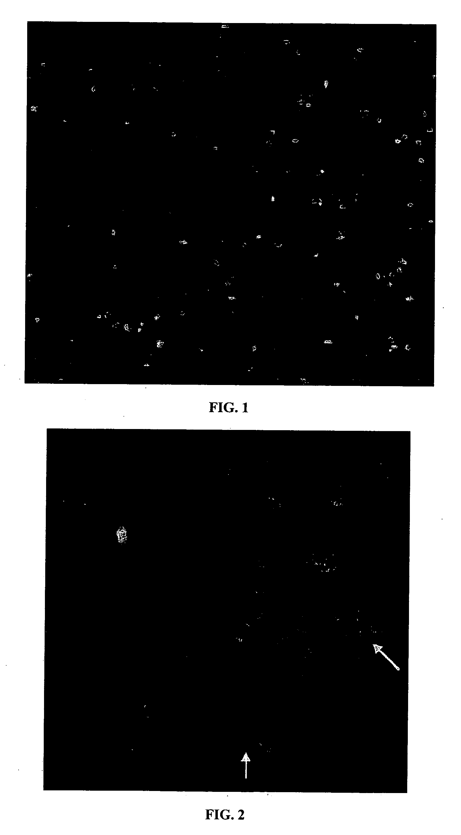
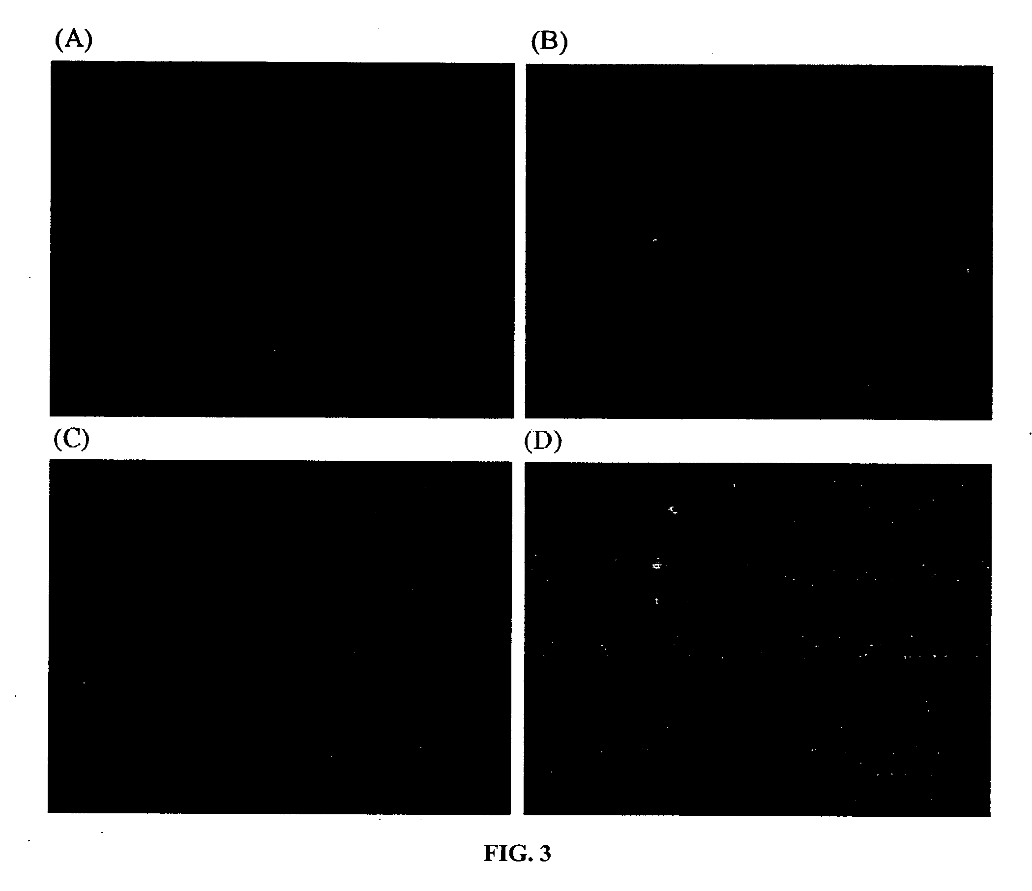
[0017] The present invention relates to a method for isolating and separating a target chromosome from a cellular sample. The method comprises the steps of (1) obtaining a cellular sample having a plurality of chromosomes, including the target chromosome; (2) arresting the cellular sample in metaphase; (3) extracting the plurality of chromosomes, including the target chromosome from the cellular sample; (4) labeling the target chromosome with a nucleic acid probe having a fluorescent reporter group to form a labeled target chromosome; (5) contacting the labeled target chromosome in suspension with an antibody against the fluorescent reporter group, with the antibody covalently linked via a linker to a magnetic bead; and (6) exposing the suspension to a magnetic field to separate the target chromosome from the plurality of chromosomes. In the present invention, the extraction step generally involves lysing the cellular sample with a hypotonic solution to form a suspension which conta...


PUM

PropertyMeasurementUnit
particle sizeaaaaaaaaaa
diameteraaaaaaaaaa

Abstract

A method for separating and isolating a target chromosome from a cellular sample by using a DNA probe with a fluorescein tag. Magnetic beads bound to anti-fluorescein isothiocyanate antibody were reacted with the fluorescently labeled pool of chromosomes and then separated in suspension by exposure to a magnetic field.

Description

CROSS-REFERENCE TO RELATED APPLICATIONS [0001] This application is based on and claims priority to U.S. Provisional Application Ser. No. 60 / 733,155, filed on Nov. 3, 2006, which is incorporated herein by reference.STATEMENT REGARDING FEDERALLY SPONSORED RESEARCH OR DEVELOPMENT [0002] Not applicable. BACKGROUND OF THE INVENTION [0003] Chromatin structure is believed to play a pivotal role in regulating genes, in addition to its function in packaging the DNA into the nucleus. The interest and effort previously devoted to exploring chromatin structure and its influence on genetic control have increased dramatically, with the rapid evolution of proteomics research after the deciphering of the human genome. Even though a single eukaryotic cell may contain thousands of genes, only those that are required for the function of a particular cell are expressed. The others are repressed by some regulatory mechanism. Another aspect of regulation is the underlying mechanism of molecular imprints,...

Claims


Application Information

Patent Timeline
Patent Type & Authority Applications(United States)
IPC IPC(8): C12Q1/68C07H21/04C12N1/08
CPCC12N15/1013C12Q1/6806C12Q2563/143
Inventor WILSON, GEORGE S.VITHARANA, SAMADHI NIPUNIKA
Owner UNIVERSITY OF KANSAS
Learn More