
Method for freezing, thawing and transplantation of viable cartilage

a cartilage and viable technology, applied in the field of freezing, thawing and transplantation of viable cartilage, can solve the problems of long time-consuming and laborious, low self-repair ability, and limited cartilage transplantation to fresh grafts, and achieve the effect of increasing the rate of warming

Inactive Publication Date: 2007-04-05
CORE DYNAMICS
View PDF55 Cites 25 Cited by
  • Summary
  • Abstract
  • Description
  • Claims
  • Application Information

AI Technical Summary

Benefits of technology

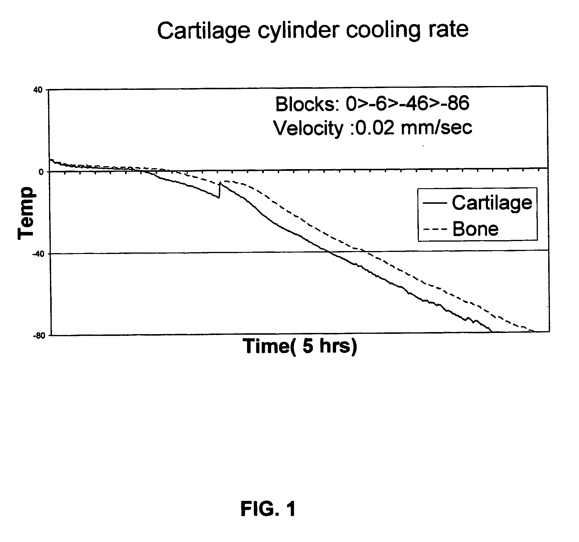
[0045] Optionally the second warming step (d) may be at a rate sufficiently slow to minimize fracture of said viable cartilage.
[0059] The warming rate of step (ii) may be the same as, or different than, that of step (iii). In fact, the warming of step (ii) may be executed at a slow rate so as to minimize the chances of fracture of said viable cartilage. Optionally, each of said steps (ii) and / or (iii) may comprise more than one warming rate.
[0066] Preferably the temperature of the environment in step (ii) would be even higher, such as 22° C. or more, 37° C. or more, or even 50° C. or 70° C. or more. The above steps are expected to be capable of increasing the rate of warming (and thus reducing the period when recrystalization may occur) since once the receptacle is removed, heat exchange may occur directly between the frozen cartilage and the solution in which it was frozen, without the receptacle being a potentially insulating barrier, without spending energy to warm the receptacle itself.
[0067] According to one option, the removal of the frozen cartilage is done by pulling it out of the receptacle at a temperature sufficiently high to allow separation of the frozen cartilage from the receptacle without significantly damaging the structure of the receptacle and / or tissue, e.g. ca. −20° C.
[0076] As detailed above, the present invention provides frozen viable cartilage. Such cartilage may remain viable for an extended period of time (practically—indefinitely), and may thus be banked. Storage and banking may be done in any method known in the art provided that the temperature of the frozen viable cartilage would not be substantially increased in an uncontrolled manner and that it would not be prematurely thawed. Any cold chamber having a temperature that is −80° C. or below, preferably −130° C. and below (e.g. liquid nitrogen vapor) or even −196° C. and below (such as liquid nitrogen) would suffice. This can allow useful or necessary tests to be performed before the tissue is transplanted (e.g. screening for infectious diseases). Banking may enable doctors to choose a graft from a larger reservoir of tissue and thus allow better donor-recipient matching (e.g. size, diameter, source and target site, shape).

Problems solved by technology

Unfortunately articular cartilage has low self-repair ability and therefore defects are prone to cause abnormal joint biomechanics, leading in the long run to degenerative changes.
Currently, cartilage transplantation is limited to fresh grafts.
This does not normally allow sufficient time to test the donated tissue for undesired agents or traits such as transmittable diseases.
It also reduces the chances of finding the best donor-recipient match.
Isolated chondrocytes (i.e. not within cartilage ECM) have been successfully cryopreserved, [1] but so far cartilage cryopreservation and control rates of freezing and thawing did not achieve an acceptable degree of cartilage viability [3].
However, it is commonly accepted that this cannot be achieved and the same assignee clearly accepts (in a later patent) that “Osteoarticular allografts, however, have not typically been used because osteoarticular cartilage cells do not survive the freezing or cryopreservation process” [6].
Nevertheless there was considerable variability in the cells' survival rates within the experimental group and the mean cell recovery was not appreciably improved.
One of the drawbacks of this method is that the added cryoprotectants may be hazardous and / or toxic and must be removed (or at least significantly diluted) upon melting.

Method used


Image

  • Method for freezing, thawing and transplantation of viable cartilage
  • Method for freezing, thawing and transplantation of viable cartilage
  • Method for freezing, thawing and transplantation of viable cartilage

Examples


Embodiment Construction

[0081] In the following, all materials were purchased from Sigma St. Louis, USA unless specified otherwise.

1—Freezing of Osteochondral Cylinders from Sheep.

[0082] Fresh cadaver sheep legs were purchased from a slaughter house (Holon Slaughter house, Israel), and all manipulations of tissue samples were done in a sterile manner. Osteochondral cylinders, 12 mm in diameter, were drilled from sheep knee chondyle using a power surgery drill (Imex, Veterinary Inc. Texas, USA). Harvested cylinders were maintained in a buffered physiological solution containing 0.9% NaCl (Sigma, St. Louis, USA) and 3% antibiotics (Penicilin / Streptomycin / Nystatin, Biological Industries, Beit Haemek, Israel) until completion of harvesting.

[0083] 10 ml cryopreservation solution (comprising nutrient mixture F-12 (HAM), 1.78M Ethylene Glycol and 1% antibiotic (penicilin / streptomycin / nystatin)) was put in a conventional 16 mm (in diameter) glass tube. The harvested cylinders were inserted into the tubes with ...


PUM


Abstract

Disclosed is a method for providing a patient having impaired cartilage in an organ at a target site, with corresponding viable cartilage, possibly osteochondral cartilage. The method comprises freezing the cartilage by cooling it at a cooling rate of 0.01° C. / min to 3° C. / min. Thawing of the cartilage may be by warming it to a temperature that is at least substantially equal to the melting temperature of the solution in which it was frozen, at a rate sufficiently high to minimize recrystalization. The thawed viable cartilage may then be grafted into the target site. Also disclosed are frozen viable cartilage and thawed viable cartilage.

Description

FIELD OF THE INVENTION [0001] The present invention relates to freezing, thawing and transplantation of viable cartilage. LIST OF REFERENCES [0002] The following references are brought to facilitate description of the background of the present invention, and should not be construed as limiting the patentability of the invention: [0003] 1. Muldrew, K. et al., Cryobiology 43, 260-267 (2001); [0004] 2. Higgs, G. B. and Boland, A. L, Proceedings of the International cartilage Repair Society's Second Symposium, November 1998; [0005] 3. McGoveran, B. M. et al., The Journal of Knee Surgery, vol. 15, No. 2 Spring 2002; [0006] 4. Williams, S K. et al, The Journal of Bone and Joint Surgery (American), 85:2111-2120 (2003). [0007] 5. U.S. Pat. No. 5,131,850 to Kelvin G. M.; [0008] 6. U.S. Pat. No. 6,488,033 to Cerundolo D. G.; [0009] 7. U.S. Pat. No. 5,873,254 to Arav A.; [0010] 8. US patent application 20030064357 to Arav A.; [0011] 9. PCT / IL03 / 00026 to Arav A. [0012] 10. U.S. Pat. No. 6,740,4...

Claims


Application Information

Patent Timeline
IPC IPC(8): A01N1/02A61K35/32
CPCA01N1/02A01N1/0284A61K35/32
Inventor DAMARI, UDIHOLTZMAN, RIVI LEVIRZEPAKOVSKY, VICTOR
Owner CORE DYNAMICS
Learn More