
Internally vented multi-function esophageal gastric tube

a gastric tube and multi-functional technology, applied in the field of esophageal gastric tubes, can solve the problems of reducing the efficiency of stethoscopes, and increasing the likelihood of significant gastric contents in patients, so as to relieve excessive negative pressure

Inactive Publication Date: 2007-03-15
GIROUX GUY M +1
View PDF13 Cites 18 Cited by
  • Summary
  • Abstract
  • Description
  • Claims
  • Application Information

AI Technical Summary

Benefits of technology

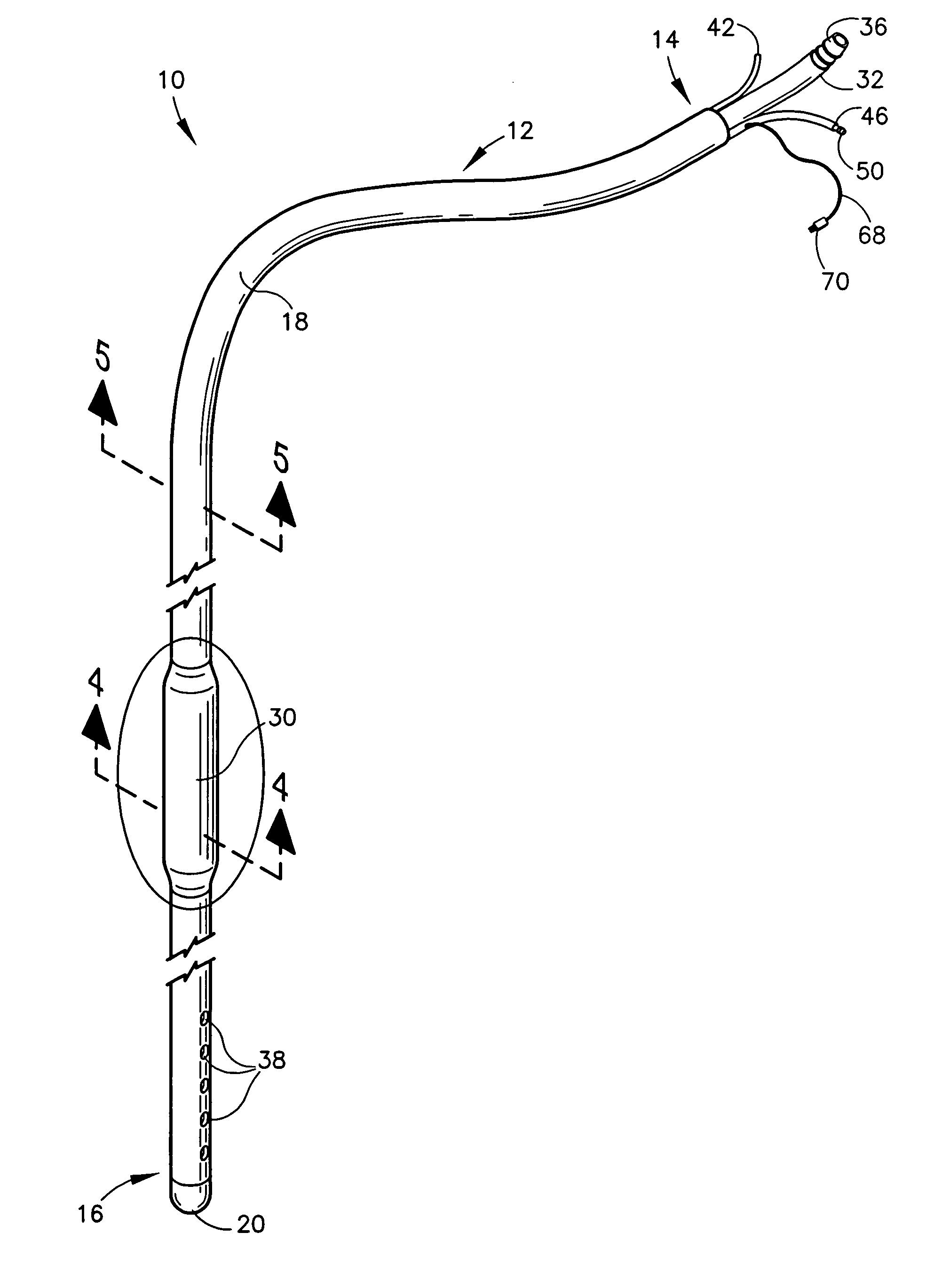
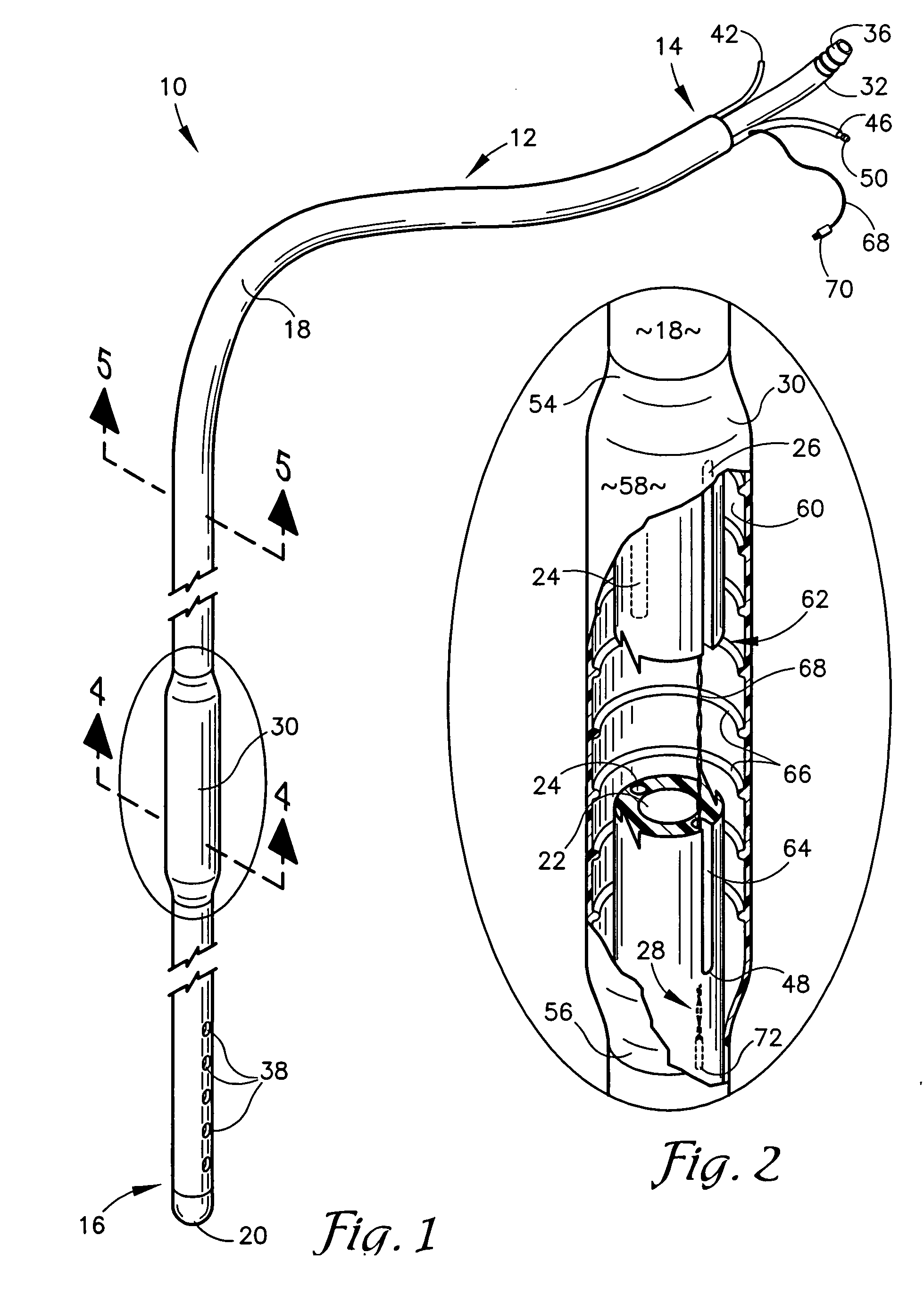
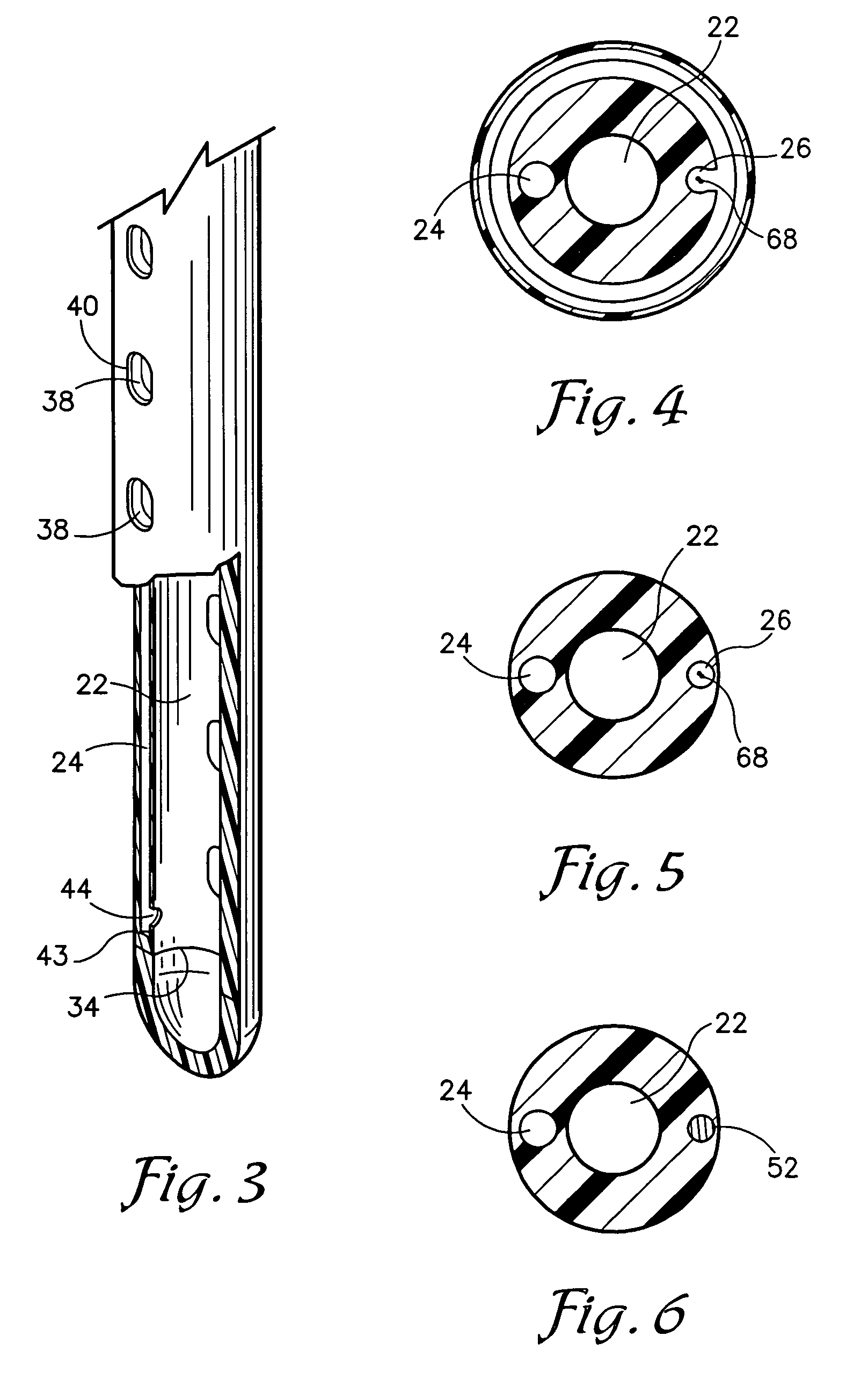
[0012] The present invention provides a greatly improved esophageal gastric tube having an open proximal end, a closed distal end, and lumens for suction, venting and auscultation. A stethoscope membrane is supported in spaced relation surrounding the tubular sidewall to form a sound chamber. The stethoscope membrane is located for positioning in the mediastinum area of a patient when the tube distal end is inserted into the patient's stomach. The stethoscope lumen extends from the proximal end of the gastric tube to at least the stethoscope membrane for transmitting body sounds. A portion of the tubular sidewall includes a longitudinal slot communicating the sound chamber with the stethoscope lumen, enabling transmission of the patient's heart and lung sounds through the stethoscope membrane and into the lumen for auscultation through acoustical earpieces connected at the proximal end of the lumen. The inner surface of the membrane includes a series of stiffening structures such as circumferential ribs for stiffening the membrane to maintain a preselected spaced relation between the inner surface of the membrane and the slot, so that the membrane will not occlude the slot and will conduct sounds around the perimeter of the tube despite compression of the tube against adjacent body tissues. A thermistor device is positioned distal of the stethoscope lumen for measuring the core temperature of the patient. The suction and vent lumens extend from the proximal end of the gastric tube to its distal end. The distal sidewall of the gastric tube is apertured through to the suction lumen to enable decompression of the stomach and suction drainage of the contents. The vent lumen extends distally to the tip area of the gastric tube, and an internal distal opening permits passage of air from the vent lumen to the suction lumen and serves as a suction break for relieving excessive negative pressure in the stomach of the patient.

Problems solved by technology

Either type of contact pressure on the membrane sleeve serves to compromise the efficiency of the stethoscope.
These conventional esophageal stethoscope devices are not well-suited to remain in place for an extended period following conclusion of a surgical procedure, and are generally removed upon conclusion of the procedure.
Emergency surgery, patient non-compliance, and the existence of certain medical conditions, such as diabetes, bowel obstruction, and obesity may increase the likelihood that the patient will have significant gastric contents that may be aspirated into the lungs during or following the surgical procedure.
A nasogastric tube may be used in conjunction with an esophageal stethoscope, however this results in crowding of the esophagus.
Most of these devices are not effectively vented, either because they do not include vent structure, or because such structure is not effective.
Attempts to include vent openings in the catheter sidewall of gastric suction devices have not generally been successful because the external apertures are subject to failure when occluded by contact with the interior wall of the stomach.
Prolonged continuous or repeated intermittent suctioning without adequate venting causes approximation of the gastric mucosa over these margins, resulting in abrasion and eventual erosion of the stomach lining by the vacuum outlet openings of the tube.
However, such external vent openings remain subject to occlusion and they fail to provide effective venting in the distal area of the tube where the suction openings are located.
Although both esophageal stethoscopes and nasogastric tubes are frequently employed, introduction of such tubes is associated with risk of injury to the tissues of the nasopharynx, hypopharynx, esophagus, larynx and associated tissues.

Method used


Image

  • Internally vented multi-function esophageal gastric tube
  • Internally vented multi-function esophageal gastric tube
  • Internally vented multi-function esophageal gastric tube

Examples


Embodiment Construction

[0021] As required, detailed embodiments of the present invention are disclosed herein; however, it is to be understood that the disclosed embodiments are merely exemplary of the invention, which may be embodied in various forms. Therefore, specific structural and functional details disclosed herein are not to be interpreted as limiting, but merely as a basis for the claims and as a representative basis for teaching one skilled in the art to variously employ the present invention in virtually any appropriately detailed structure.

[0022] Referring now to the drawing figures, the reference numeral 10 refers to an esophageal gastric tube apparatus in accordance with the invention. As best shown in FIGS. 1 and 3, the gastric tube 10 includes an elongated catheter or tube 12, having a proximal end 14 and a distal end 16 with a connecting sidewall 18. The proximal end 12 is generally open, whereas the distal end 16 is sealed or closed to form a tip 20.

[0023] The tube 12 includes a first ...


PUM

PropertyMeasurementUnit
Angleaaaaaaaaaa
Temperatureaaaaaaaaaa
Pressureaaaaaaaaaa

Abstract

An esophageal gastric tube includes lumens for suction, venting and auscultation. The auscultation lumen serves as a conduit for electrical conductors to a thermistor positioned distally of the lumen for measuring core temperature without occlusion of the lumen. A stethoscope membrane encircles the tube, forming a sound chamber. Suction and vent lumens extend from the proximal tube end to the distal tip, while the stethoscope lumen terminates at the sound chamber distal end, for reducing suction noise interference with auscultation. A longitudinal slot in the tubular sidewall enables effective communication between the sound chamber and the stethoscope lumen. The stethoscope membrane includes internal circumferential ribs preventing occlusion of the slot and permitting 360° auscultation. The tube distal sidewall is apertured into the suction lumen, enabling suctioning of the gastric contents. Vent and suction lumens communicate within the distal end of the tube to provide an internal vent for relieving negative stomach pressure.

Description

BACKGROUND OF THE INVENTION [0001] The present invention is broadly concerned with a tubular apparatus for insertion through the nose or mouth and into the stomach of a patient for simultaneously detecting heart and lung sounds, monitoring core body temperature, and providing suction drainage and gastric pressure equalization. More particularly, it is concerned with a multiluminate esophageal gastric tube having a temperature sensor and stethoscope, a suction lumen and a vent lumen with an internal distal opening. [0002] In surgical procedures performed on patients under general anesthesia, an esophageal stethoscope is generally introduced into the esophagus in order to enable continuous monitoring of the patient's heart and breath sounds. The device may also include thermistor structure for monitoring the patient's core body temperature. Such monitoring is necessary in order to promptly detect any variations from the normal which may occur during the procedure, as well as to guide ...

Claims


Application Information

Patent Timeline
IPC IPC(8): A61B5/02
CPCA61B1/273A61B5/01A61M2025/0037A61B7/023A61M25/0045A61B5/4233
Inventor GIROUX, GUY M.GLASGOW, JEFFERY W.
Owner GIROUX GUY M
Learn More