Hematological assay and kit
a kit and assay technology, applied in the field of assays and kits, can solve the problems of inability to perform standard laboratory analyses, inability to use defibrinated samples for the standard analysis of the coagulation system, and inability to detect fibrin polymerisation in time, etc., and achieve the effect of convenient detection
- Summary
- Abstract
- Description
- Claims
- Application Information
AI Technical Summary
Benefits of technology
Problems solved by technology
Method used
Image
Examples
example 1
[0100] For the assessment of the inventive assay the following solutions were prepared:
[0101] solution 1 an aqueous solution containing CaCl2 (25 mM), a chromogenic substrate (250 μM H-D-CHG-Ala-Arg-pNa·2AcOH, Pentapharm, Basle) and a fibrin polymerisation inhibitor (5 mg / ml H-Gly-Pro-Arg-Pro-OH AcOH, Pentapharm, Basle).
[0102] solution 2 an aqueous solution containing a contact activator (aluminium silicate 5 g / l, Sigma, St. Louis, USA) and phospholipids (derived from rabbit brain cephalin, 50 μg / ml, Pentapharm, Basle).
[0103] Platelet poor plasma was prepared from venous citrated blood by centrifugation (20 min. at 1500 g).
[0104] The procedure was performed as follows:
[0105] 50 μl platelet poor plasma was mixed with 50 μl solution 2 and incubated for 180 seconds. 50 μl solution 1 was then added and detection of the optical density carried out at 405 nm over 400 seconds.
[0106] This procedure is generally followed with specified variations in the (inventive) examples which follo...
example 2
[0107] The procedure was repeated with different substrate concentrations of the substrate
[0108] H-D-CHG-Ala-Arg-pNa·2AcOH (KM 15,9 μM, Pentapharm, Basle)to obtain the graph of FIG. 5.
[0109] From the reaction curves the first derivative was calculated by determining the rise of optical density over each 4 seconds as already described. The first derivative is plotted in FIG. 6.
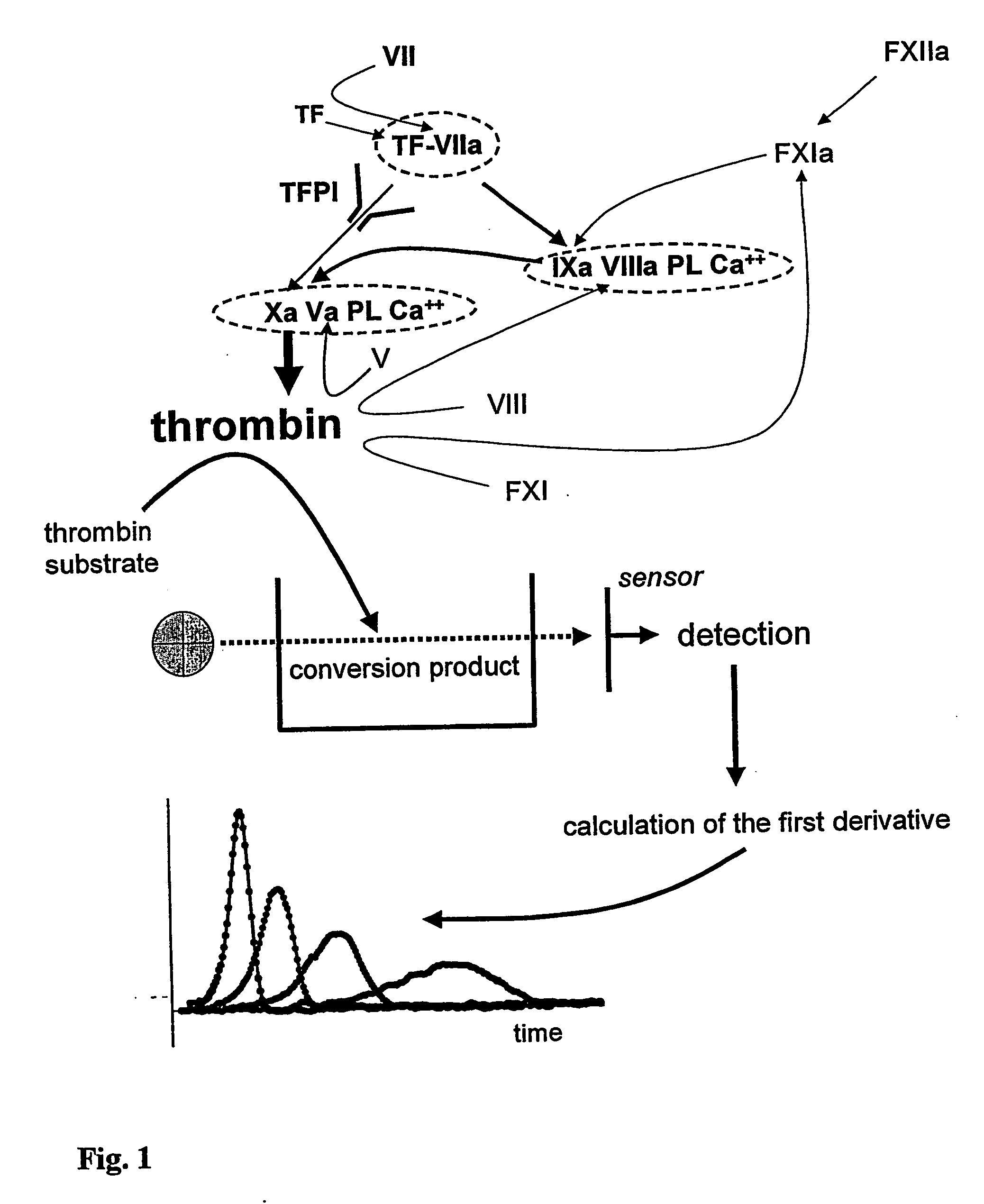
[0110]FIG. 5 demonstrates the thrombin formation in the same plasma as in FIGS. 3 and 4 detected with the method according to the--present invention. Using the inventive method a much stronger (20 times stronger) optical signal is generated. The rise of the optical density stops when the substrate is consumed, typically 1-2 min. after the onset of thrombin formation. FIG. 5 and FIG. 6 show the effect of rising concentrations of the substrate on the detection properties. Rising concentrations of the substrate lead to a rising optical signal. However already the lowest concentrations tested are sufficient for ...
example 3
[0111] The procedure of Example 1 was repeated using different concentrations of the activator of the plasmatic coagulation system on the assessment of the thrombin formation using the inventive assay. Recombinant Tissue Factor (Instrumentation Laboratory, Kirchheim, Germany) was serially diluted and used as the activator in solution 2. The results of the optical densities and first derivatives are shown respectively in FIG. 7 and 8. The numbers in the diagrams show the concentration of recombinant Tissue Factor [ng / ml sample]. Thus the assay can be adapted as required depending on the activation procedure applied.
PUM
 Abstract
Description
Claims
Application Information
 - R&D
- Intellectual Property
- Life Sciences
- Materials
- Tech Scout
- Unparalleled Data Quality
- Higher Quality Content
- 60% Fewer Hallucinations
Browse by: Latest US Patents, China's latest patents, Technical Efficacy Thesaurus, Application Domain, Technology Topic, Popular Technical Reports.
© 2025 PatSnap. All rights reserved.Legal|Privacy policy|Modern Slavery Act Transparency Statement|Sitemap|About US| Contact US: help@patsnap.com


