
Ultrasonic device and method for treating stones within the body

a technology of ultrasonic devices and ureters, which is applied in the field of ultrasonic devices and methods for treating stones within the body, can solve the problems of difficult image of ureter stones in some portions of the ureter, the inability of ultrasound to substantially affect patient tissue, and the limitations of the use of extra-corporeal shock wave lithotripsy (eswl) techniques

Inactive Publication Date: 2006-08-17
ETHICON ENDO SURGERY INC
View PDF19 Cites 86 Cited by
  • Summary
  • Abstract
  • Description
  • Claims
  • Application Information

AI Technical Summary

Benefits of technology

[0018] The present invention is useful in open or endoscopic surgeries as well as robotic-assisted surgeries.
[0019] Further features and advantages of the present invention will become readily apparent from the following detailed description, the accompanying drawings, and the appended claims.

Problems solved by technology

However, focused medical-treatment ultrasound will not substantially medically affect patient tissue outside the focal zone such as patient tissue located between the source and the focal zone.
For stones in the ureter, there are substantial limitations to the use of extra-corporeal shock wave lithotripsy (ESWL) techniques.
Ureter stones in some portions of the ureter can be difficult to image because of interfering structure in the body.
Similarly extra-corporeal techniques may not work for heavy patients at or above 300 pounds.
Near this weight and above, it may not be possible to focus the ultrasonic energy to reach a stone.
ESWL can also be complicated in cases where a patient has a pre-existing pulmonary or cardiac problem as shock waves can cause dysrhythmias.
Another limitation of ESWL can be on larger stones and persistent steinstrasse.
Extra-corporeal shock wave techniques may also not be effective for some stone compositions.
ESWL may not work well for stones of calcium monohydrate, calcium phosphate and cystine.
Problems with this technique include size and rigidity of the probe which generally limit applications to stones in the lower portion of the ureter.
The technique can also cause unpredicted movement of the stone, which can lead to tissue damage.
Potential problems with electrohydraulic lithotripsy include heating, unpredictable stone movement and potential tissue damage to the ureter.
Problems with laser lithotripsy can include tissue damage, and heat.
An additional consideration is that the laser units and fibers can be expensive.
Another problem with prior art lithotripsy using internal imaging is with the ability to image the stone.
Often the visual field using fiber optic scopes can be obscured and there is no depth of field.
With poor imaging, it is possible to actually push the stone up the ureter with the probe because an operator cannot see the stone.
Such unplanned movement of the stone is undesirable as it can lead to chasing a moving target or worse injury to the wall of the ureter.
Another problem with transurethral lithotripsy has been that these procedures have several risk factors that typically require the procedure to take place on an inpatient basis and with the use of a full surgical suite.
These risk factors include the risks associated with the use of general anesthetic, the risk of perforations to the ureter wall, and the need to be able to place a stent.
Further, current techniques typically require a fluoroscope to perform the initial imaging and fluoroscopes are an expensive piece of medical equipment typically only available within a surgical suite.

Method used


Image

  • Ultrasonic device and method for treating stones within the body
  • Ultrasonic device and method for treating stones within the body
  • Ultrasonic device and method for treating stones within the body

Examples


Embodiment Construction

[0024] Before explaining the present invention in detail, it should be noted that the invention is not limited in its application or use to the details of construction and arrangement of parts illustrated in the accompanying drawings and description. The illustrative embodiments of the invention may be implemented or incorporated in other embodiments, variations and modifications, and may be practiced or carried out in various ways. Furthermore, unless otherwise indicated, the terms and expressions employed herein have been chosen for the purpose of describing the illustrative embodiments of the present invention for the convenience of the reader and are not for the purpose of limiting the invention.

[0025] The features of the invention are set forth with particularity in the appended claims. The invention itself, however, both as to organization and methods of operation, together with further objects and advantages thereof, may best be understood by reference to the following descr...


PUM


Abstract

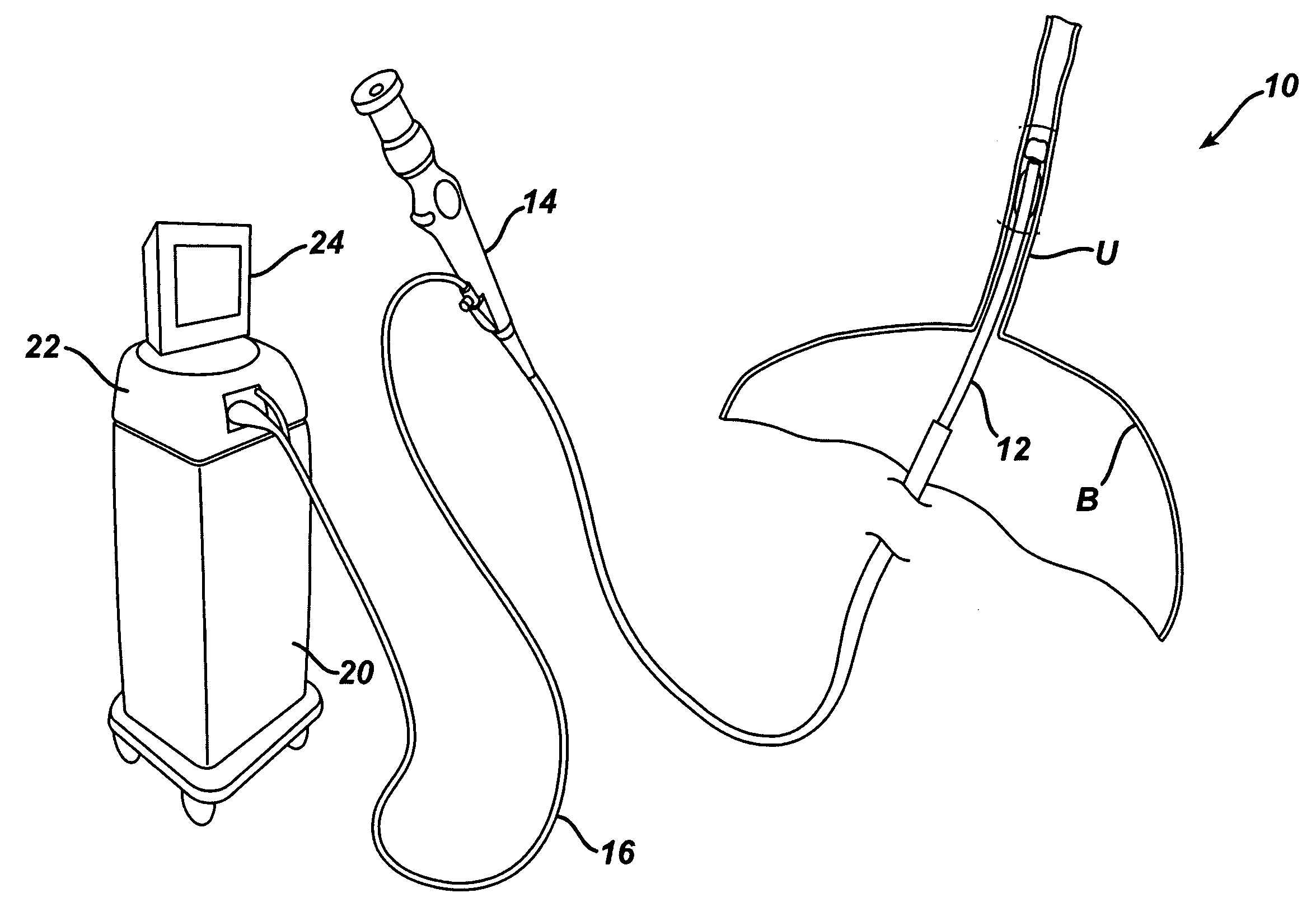
A system and method to be used in ultrasonic lithotripsy of a stone in a ureter, the system including a catheter having a probe tip capable of transmitting and receiving ultrasonic energy. The catheter can include an inflatable balloon adjacent to the probe tip, the balloon capable of pooling some urine in the ureter to be used as an ultrasonic transmission media. The ultrasonic probe is connected to a source of energy capable of driving the probe tip to deliver ultrasonic energy of a high frequency and relatively low energy to image a stone. Then the probe can be connected to a source of energy capable of driving the probe to deliver a low frequency, high energy ultrasonic to disintegrate the stone.

Description

CROSS REFERENCE TO RELATED APPLICATION [0001] The present application claims the priority benefit of U.S. provisional patent application Ser. No. 60 / 632,016, filed on Dec. 1, 2004, which is incorporated herein by reference in its entirety. [0002] This application is related to U.S. patent application Ser. No. ______, attorney docket END-5485USNP, filed concurrently herewith.FIELD OF THE INVENTION [0003] The present invention relates to apparatus and method to ultrasonically image and break apart a stone in the ureter. BACKGROUND OF THE INVENTION [0004] Known ultrasound medical systems and methods include using ultrasound imaging of patients to identify patient tissue for medical treatment and include using ultrasound to medically destroy identified patient tissue by heating the tissue. Imaging is done at lower power and medical treatment is done at higher power. Low power imaging ultrasound will not medically affect patient tissue. High power medical-treatment ultrasound, when focus...

Claims


Application Information

Patent Timeline
Patent Type & Authority Applications(United States)
IPC IPC(8): A61H1/00
CPCA61B17/22012A61B17/2202A61B2017/00022A61B2017/00137A61B2017/22067A61B2019/5276A61B2019/528A61B2090/378A61B2090/3784
Inventor GILL, ROBERT P.VOEGELE, JAMES W.WEISENBURGH, WILLIAM BRUCE II
Owner ETHICON ENDO SURGERY INC
Learn More