Detection of Trichomonas

a technology of trichomonas and diagnostic devices, applied in the field of diagnostic devices and methods for detecting trichomonas infections, can solve problems such as severe complications, and achieve the effect of facilitating detection of infections

Inactive Publication Date: 2006-04-27
KIMBERLY-CLARK WORLDWIDE INC
View PDF5 Cites 8 Cited by
  • Summary
  • Abstract
  • Description
  • Claims
  • Application Information

AI Technical Summary

Benefits of technology

[0007] One aspect of the invention is a diagnostic device that includes a core and a multitude of reporter enzymes, wherein each reporter enzyme is directly or indirectly linked to the core by a linker, and wherein the linker can be cleaved by a Trichomonas-specific enzyme after insertion of the device in a mammalian vagina. The core can be surrounded by membrane to which the reporter enzymes are linked. The reporter enzymes can be present in one zone of the core (this may facilitate detection of the infection). The Trichomonas-specific enzyme can be a Trichomonas vaginalis enzyme or a Trichomonas foetus enzyme. The reporter enzymes can be proteinases, phospholipase A2-like lytic enzymes, or neuraminidases. In some embodiments, the reporter enzymes are cysteine proteinases. The reporter enzymes can be proteinases that cleave a linker with a cleavage site that includes Arg-Arg, Phe-Arg, Val-Leu-Lys, Ala-Phe-Lys, Pro-Phe-Arg, or Leu-Val-Tyr. In another embodiment, the reporter enzymes are phospholipase A2-like enzymes. Such phospholipase A2-like enzymes can cleave a linker with a cleavage site that includes a phosphoglyceride. The phospholipase A2-like enzymes can also cleave an alkylene linker with a cleavage site comprising:

Problems solved by technology

The infection recently has been associated with severe complications, including infertility.
However, given the prevalence of Trichomonas infections and their potential harm, faster, simpler methods for detecting Trichomonas infections are needed.

Method used


Image

  • Detection of Trichomonas
  • Detection of Trichomonas

Examples


Embodiment Construction

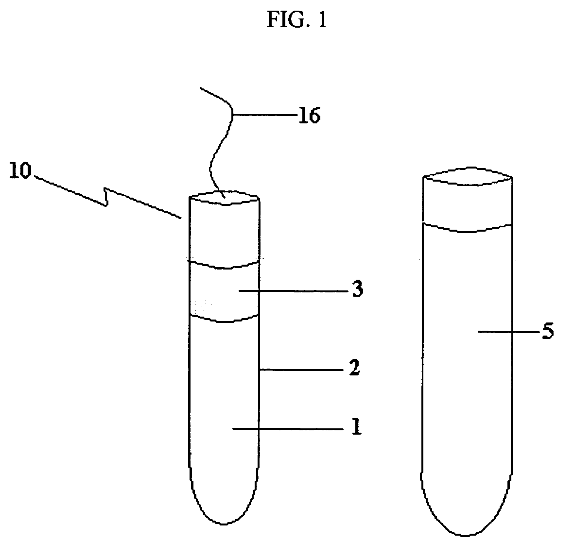
[0015] The invention relates to diagnostic devices and methods for detecting vaginal infections, for example, Trichomonas infections. The diagnostic devices of the invention include a vaginal insert or tampon having a solid support core material with at least one antibody specific for an antigen of an infective agent or at least one reporter enzyme attached to the core material or to a membrane covering the core material. The reporter enzyme is attached to the core or membrane by a linker that can be cleaved by an enzyme specific to an infective agent. The infective agent-specific antibodies or reporter enzymes can also be bound to and be within the material of the solid support core. The methods of the invention involve inserting a diagnostic device of the invention into a female mammal's vagina for a time sufficient to permit binding of infective agent-specific antigens or interaction of the reporter enzymes with the infective agent-specific enzyme. The diagnostic device can then ...


PUM

PropertyMeasurementUnit
lengthaaaaaaaaaa
sizeaaaaaaaaaa
lengthaaaaaaaaaa

Abstract

The present invention is directed to diagnostic devices and methods for detecting Trichomonas infections.

Description

FIELD OF THE INVENTION [0001] The invention relates to diagnostic devices and methods for detecting Trichomonas infections. BACKGROUND OF THE INVENTION [0002]Trichomonas vaginalis is the cause one of the most widespread sexually transmitted diseases in the world. The main pathological manifestations of a trichomonad infection in women are abdominal pain, itching, and presence of a foul-smelling discharge with abundant leukocytes Honigberg, B. M. 1978. Trichomonads of importance in human medicine, p. 275. In J. P. Kreier (ed.), Parasitic protozoa, vol. 11. Academic Press, Inc., New York, N.Y. In men, the infection is mostly asymptomatic, although it can sometimes lead to urethritis, prostatitis, and epididymitis. Krieger, J. N. 1981. Urologic aspects of trichomoniasis. Investig. Urol. 18:411-417. The infection recently has been associated with severe complications, including infertility. Moskowitz, M. O., and B. C. Mellinger. 1992. Sexually transmitted diseases and their relation to ...

Claims


Application Information

Patent Timeline
Patent Type & Authority Applications(United States)
IPC IPC(8): G01N33/569C12Q1/04C12M1/34
CPCA61B10/0045A61B2010/0006A61B2010/0074A61F13/202A61F13/2074A61F2013/00429A61F2013/8473C12Q1/04G01N33/54366G01N33/56905
Inventor WEART, ILONAYANG, SHU-PING
Owner KIMBERLY-CLARK WORLDWIDE INC
Why Eureka
Try Eureka