
Luciferyl peptide substrate

a technology of luciferin and substrate, which is applied in the field of multifunctional bioluminescent substrate preparation, can solve the problems of difficult identification of reaction and its parameters within the system, complex vivo studies, and inability to take into account the effects of in vitro reactions, and achieve the effect of facilitating localization of luciferin-agent conjugates

Inactive Publication Date: 2006-04-06
THE BOARD OF TRUSTEES OF THE LELAND STANFORD JUNIOR UNIV
View PDF2 Cites 24 Cited by
  • Summary
  • Abstract
  • Description
  • Claims
  • Application Information

AI Technical Summary

Benefits of technology

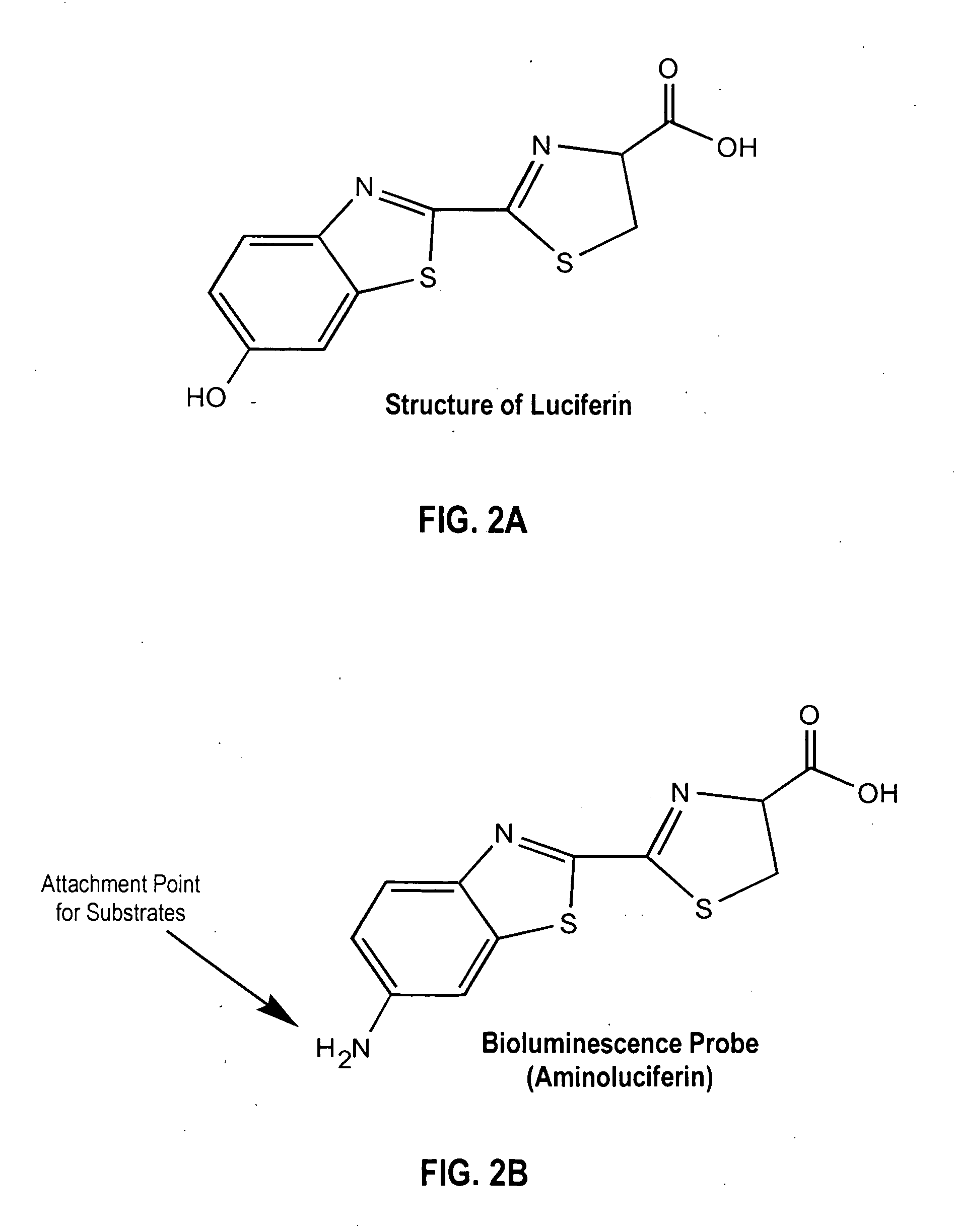
[0013] In a further embodiment of the invention, there is provided a method of delivering agents to specific sites in an animal species, including humans, comprising the steps of (a) conjugating aminoluciferin with an imaging agent to form an aminoluciferin-agent conjugate; (b) conjugating the aminoluciferin-agent conjugate with a peptide sequence that cannot penetrate cell membranes or tissue barriers, to produce an agent-luciferyl peptide substrate that will not penetrate cell membranes or tissue barriers; (c) injecting the animal species with the agent-luciferyl peptide substrate, wherein within the animal species, the peptide sequence is cleaved by a target enzyme on a target cell or tissue to reform the luciferin-agent conjugate; (d) monitoring the animal species for signals from the luciferin-agent conjugate that indicate passage of the luciferin-agent conjugate across the cell membrane or tissue barrier and retention of the luciferin-agent conjugate in cells or tissue, wherein the signal from the luciferin-agent facilitates localization of the luciferin-agent conjugate. The cell membranes include, without limitation, tumor cell membranes, neuronal membranes, and other cell membranes, and the tissue barriers include, without limitation, placental barriers and blood-brain barriers.

Problems solved by technology

On the other hand, in vitro reactions do not usually take into account the effects of the surrounding systems on the particular reaction being studied.
In vivo studies are much more complex, as multiple systems may be affecting the reaction being studied, making it difficult to identify the reaction and its parameters within the systems.
Drawbacks of using fluorescent markers include the requirement to stimulate the markers by an energy source outside the tissue or animal being studied.
This can cause scatter and low efficiency of the marker.
Interference from autofluorescence from endogenous molecules, such as hemoglobin and cytochromes, can also decrease the ability to detect such markers.

Method used


Image

  • Luciferyl peptide substrate
  • Luciferyl peptide substrate
  • Luciferyl peptide substrate

Examples


example 1

Aminoluciferyl Substrates and Enzymes for Use in in vivo Models

[0051] The efficacy of the luciferyl peptide substrates of the present invention in in vivo models may be determined by using the five enzymes and the eight aminoluciferyl peptide substrates shown in Table 2.

TABLE 2ENZYMEAMINOLUCIFERYL SUBSTRATESCathepsin B1) Bz-Arg-aLuc2) Pyr-Phe-Leu-aLuc3) Z-Arg-Arg-aLucPSA4) Ac-His-Ser-Ser-Lys-Leu-Gln-aLuc5) Ac-Ser-Lys-Leu-Gln-aLucMMP-2 and MMP-96) Ac-Gly-Pro-Leu-Gly-Ile-Ala-Gly-Gln-aLucThrombin7) Bz-Phe-Val-Arg-aLucHIV Protease8) Abz-Thr-Ile-Nle-aLuc-Phe-Gln-Arg-NH2

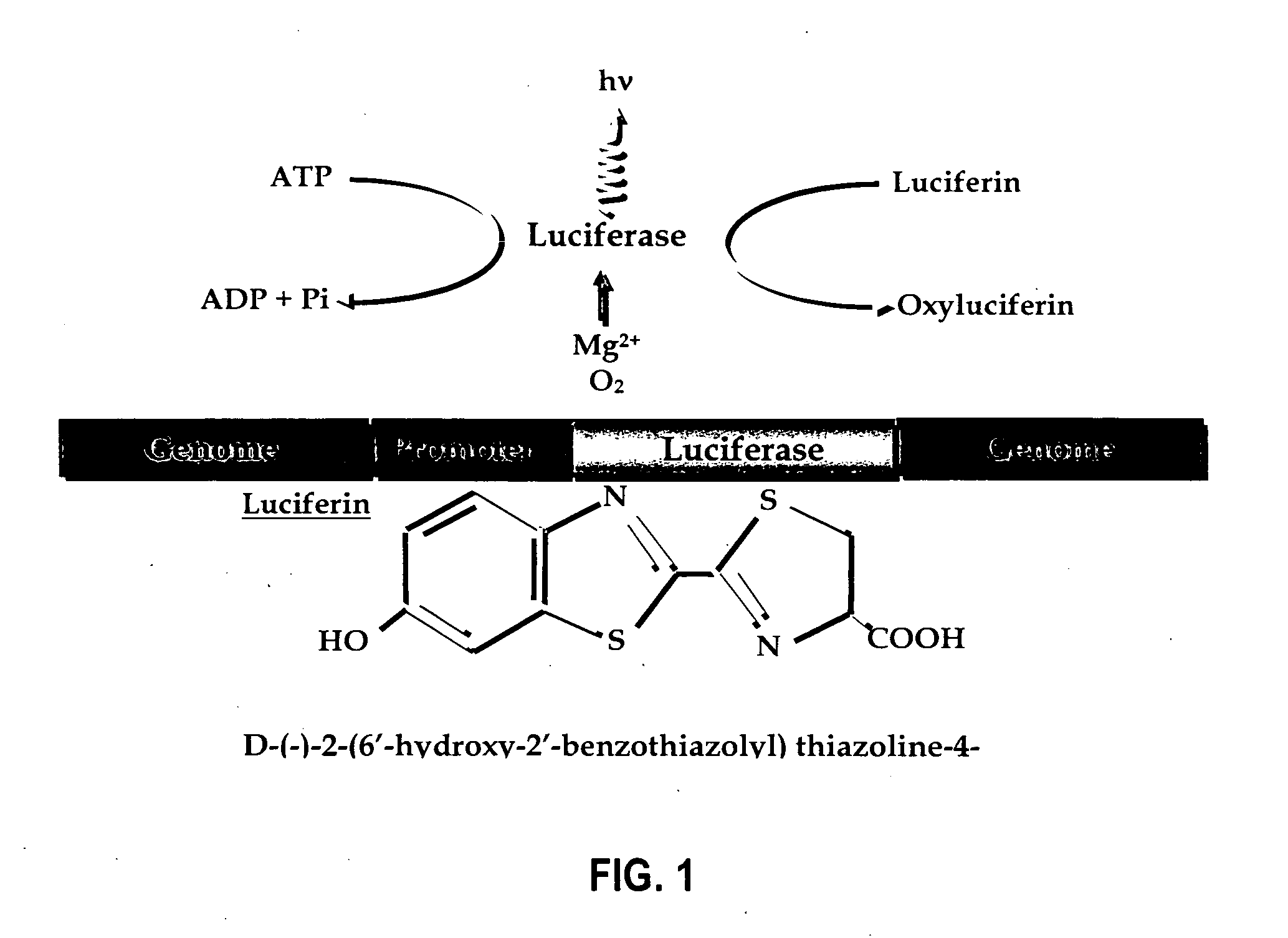
[0052] The aminoluciferyl peptide substrate identified by No. 5 in Table 2 (Ac-Ser-Lys-Leu-Gln-aLuc referred to as “SKLQ-aLuc”) was synthesized as a substrate for PSA. The sequence selected for the peptide is relatively specific for PSA over other enzymes. The amount of aminoluciferin released is quantified by its instantaneous reaction with luciferase.

example 2

The Effect of PSA-Specific Aminoluciferyl Peptide Substrates on PSA Secreting Cells—an in vitro Model

[0053] To study the relationship of the PSA-specific aminoluciferyl peptide substrates on PSA secreting cells, SKLQ-aLuc was introduced in LNCaP, a prostate cancer cell line that produces PSA, and PC3M, a prostate cancer cell line that does not produce PSA. The LNCaP cells were incubated for 5 and 19 hours, which represented two time intervals during which the cells were to synthesize PSA, and both cell lines were transfected with SKLQ-aLuc to produce luciferase. Dihydroxytestosterone (“DHT”) was used to investigate its influence on cell growth and PSA production. The cell culture media was serum-free to prevent PSA from forming complexes with various serine-protease inhibitors that are present in the serum. The results of this experiment are shown in FIG. 7, which demonstrate that the amount of aminoluciferyl peptide detected in the cells is directly proportional to the amount of P...

example 3

The Effect of PSA-Specific Aminoluciferyl Peptide Substrates on PSA Secreting Cells—an in vivo Model

[0054] The peptides were then tested in severe compromised immune deficiency (SCID) mice that bore an LNCaP tumor implanted at a subcutaneous site. FIG. 8A shows the presence of 2 week old LNCaP tumors on the right flank of the mice. The mice were injected with luciferin to localize the tumors and confirm their presence. FIG. 8B shows light emission from these tumors 2 months after injecting the PSA-specific aminoluciferyl peptide SKLQ-aLuc. The aminoluciferin released from the cleavage of the peptide by PSA is transported across the LNCaP cell membrane and reacts with luciferase to emit light. Thus, the aminoluciferyl peptide is activated by PSA and consequently can be used to target PSA producing cells in animal models.


PUM

PropertyMeasurementUnit
emission wavelengthaaaaaaaaaa
structureaaaaaaaaaa
Biological imagingaaaaaaaaaa

Abstract

Provided are luciferyl peptide substrates that are produced by attaching specifically prepared peptide conjugates to luciferin, and / or its analogs and derivatives. The luciferyl peptide substrates are incapable of penetrating cell membranes and tissue barriers. Cleavage of the peptide conjugates from the luciferyl peptide substrates releases the luciferin, which upon contact with luciferase emits photons for easy detection. The luciferyl peptide substrates may be used in assays to detect pathogens, test protease inhibitors, probe cell physiology, assess protease activity in oncogenesis, and improve specific and regulated drug delivery.

Description

TECHNICAL FIELD [0001] The invention relates generally to the field of multifunctional bioluminescent substrates. More specifically, the invention relates to the preparation of luciferyl peptide substrates from luciferin, its analogs, and derivatives combined with specifically prepared peptides, and the use of the luciferyl peptide substrates in improved assays to detect pathogens, test protease inhibitors, probe cell physiology, assess protease activity in oncogenesis, and improve specific and regulated drug delivery. BACKGROUND OF THE INVENTION [0002] Biological processes can be studied both in vitro and in vivo. In vitro methods provide the ability to isolate molecules from the complex milieu of a biological system. Thus, they provide simpler settings for studying individual reactions than can be found in vivo. On the other hand, in vitro reactions do not usually take into account the effects of the surrounding systems on the particular reaction being studied. In vivo studies are...

Claims


Application Information

Patent Timeline
Patent Type & Authority Applications(United States)
IPC IPC(8): G01N33/574C12Q1/66C07K7/06
CPCC07K5/1013C12Q1/37C12Q1/66
Inventor CONTAG, CHRISTOPHER H.SHINDE, RAJESH R.BEDNARSKI, MARK D.GUCCIONE, SAMIRA
Owner THE BOARD OF TRUSTEES OF THE LELAND STANFORD JUNIOR UNIV
Learn More