Tomographic reconstruction of transmission data in nuclear medicine studies from an array of line sources

a line source and transmission data technology, applied in the field of single photon emission computed tomography (spect) nuclear medicine studies and correction of data attenuation, can solve the problems of inability to accurately represent line integrals of radioisotopes in quantitative image values in various projections, and distortions introduce errors or artifacts,

Inactive Publication Date: 2006-03-30
SIEMENS MEDICAL SOLUTIONS USA INC
View PDF2 Cites 2 Cited by
  • Summary
  • Abstract
  • Description
  • Claims
  • Application Information

AI Technical Summary

Benefits of technology

[0013] The preferred embodiments of the present invention can significantly improve upon existing methods and / or apparatus. According to a preferred embodiment of one aspect of the invention, a new type of algorithm for μ-map reconstruction uses a statistically-based estimation of the μ-map. Such algorithm allows overlapping of line source radiation patterns at the detector, and additive inclusion of emission-to-transmission downscatter.

Problems solved by technology

When FBP reconstruction is used to reconstruct SPECT images from two-dimensional projection images obtained from a scintillation camera, some well-recognized distortions introduce errors or artifacts in the result.
As a consequence of attenuation, quantitative image values in the various projections do not accurately represent line integrals of the radioisotope distribution within the body.
However, for a very important class of studies, namely cardiac SPECT studies, the linear attenuation coefficient of the body is in fact highly non-uniform.
However, present methods suffer from certain disadvantages.
In particular, FBP does not optimally process the noise or distortion in the projection data.
FBP is not statistically based, and the conventional FBP computational algorithm is prone to “streak” artifacts predominantly oriented in the radial direction.
Another problem with existing attenuation correction methods involves the correction of transmission CT data for downscatter by subtracting estimated downscatter values from the transmission data.
Attenuation of the transmission radiation beam through a patient can be large (˜50), resulting in count-starved data.
Therefore, the scatter in this region is not the same as that in the heart region, and using a sub-cardiac region for estimation of the downscatter fraction can seriously bias the estimate, causing errors in the attenuation map (i.e., mu-map).

Method used


Image

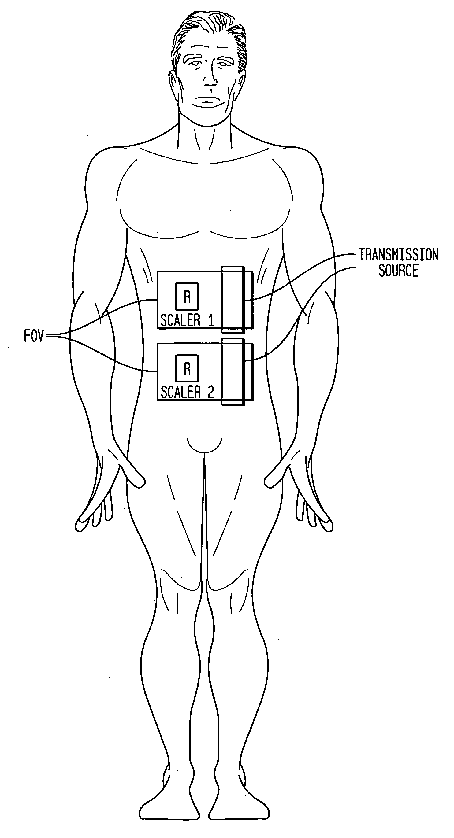
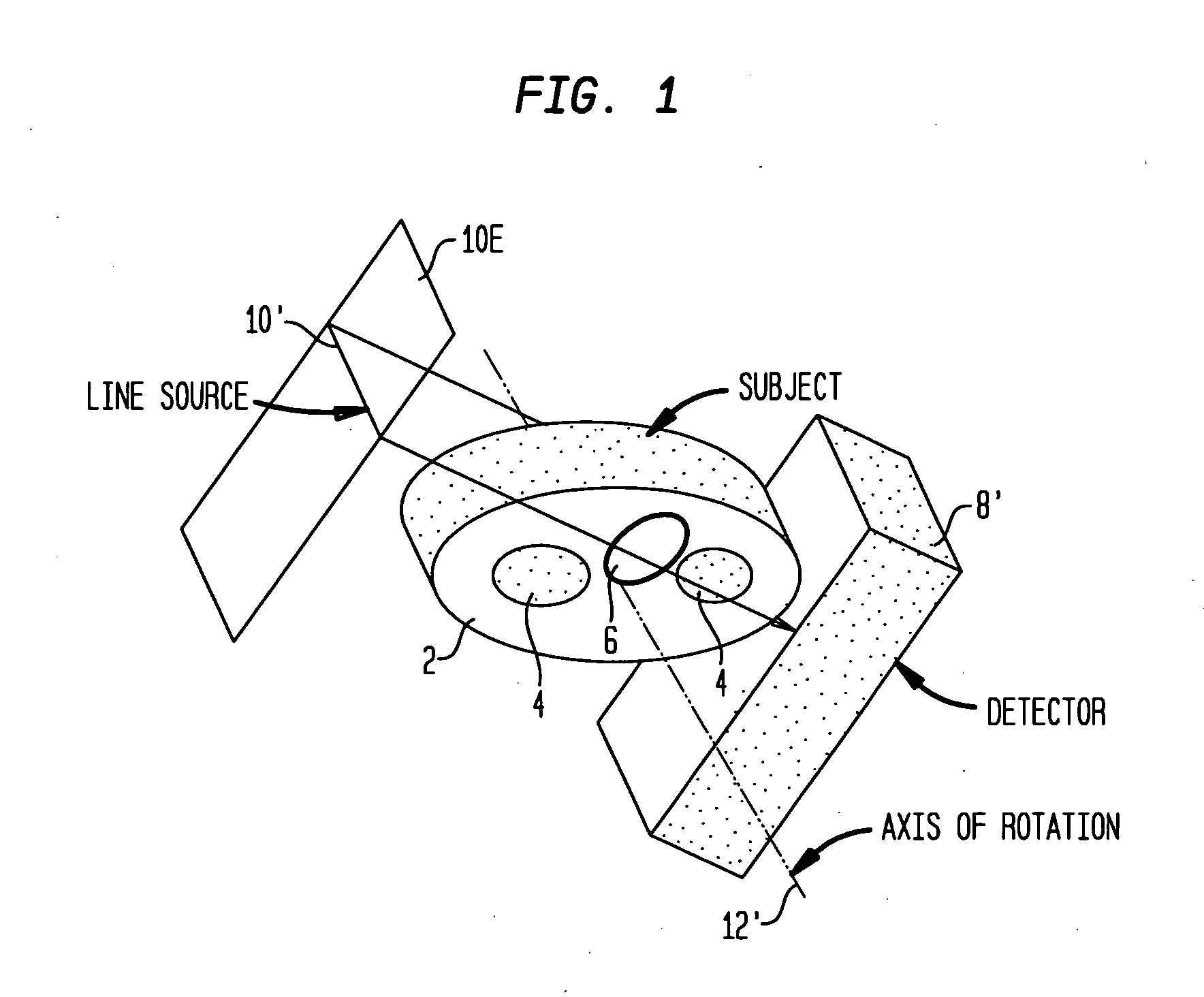
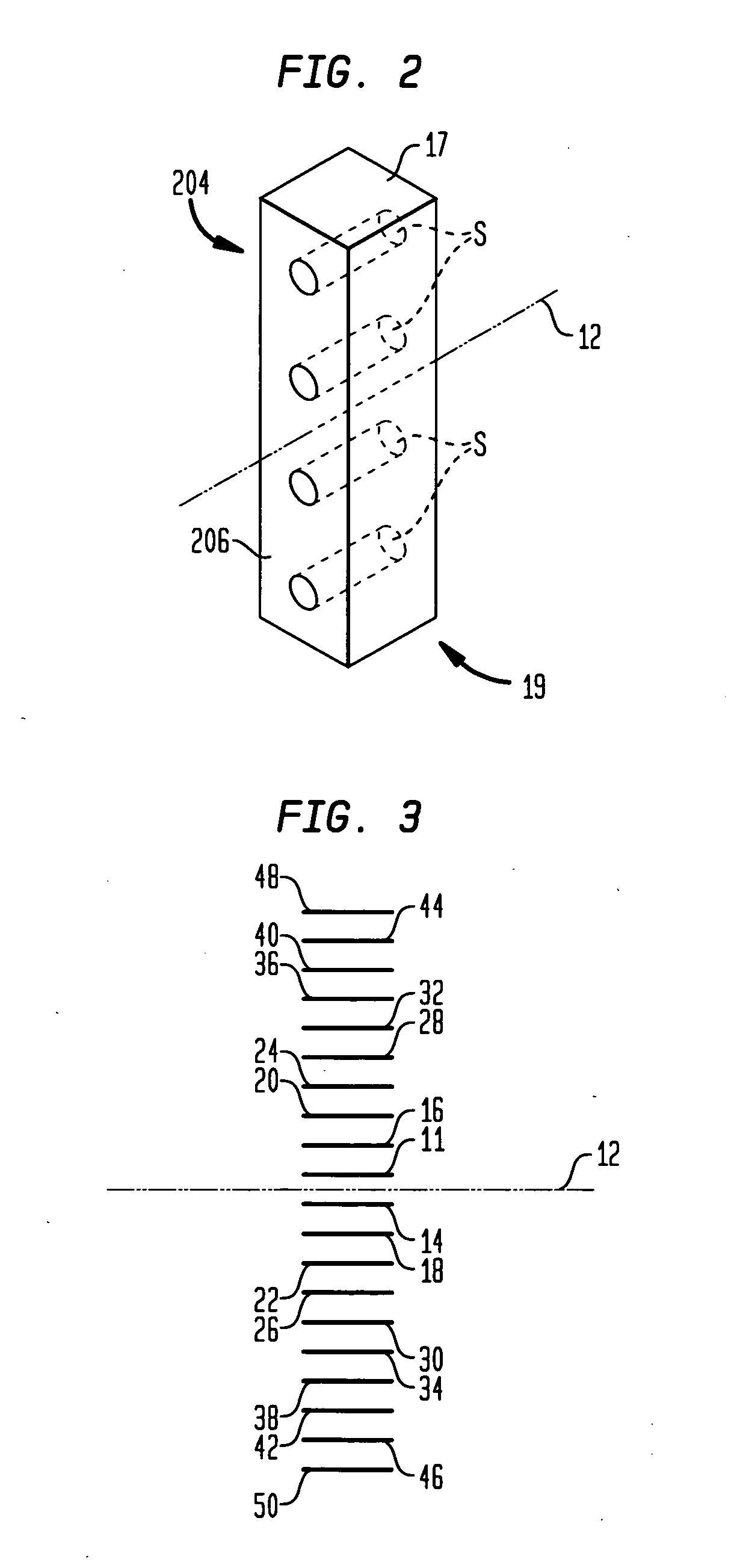
  • Tomographic reconstruction of transmission data in nuclear medicine studies from an array of line sources
  • Tomographic reconstruction of transmission data in nuclear medicine studies from an array of line sources
  • Tomographic reconstruction of transmission data in nuclear medicine studies from an array of line sources

Examples


Embodiment Construction

[0020] While the present invention may be embodied in many different forms, a number of illustrative embodiments are described herein with the understanding that the present disclosure is to be considered as providing examples of the principles of the invention and such examples are not intended to limit the invention to preferred embodiments described herein and / or illustrated herein.

Summary of Attenuation Correction Procedure and Set-Up

[0021] Before explaining the various aspects and preferred embodiments of the present invention, a brief explanation will be given of a conventional procedure for obtaining transmission CT data for attenuation correction in SPECT studies. In a SPECT study, a collimated detector is rotated to a plurality of consecutive angularly separated stationary positions around a patient. Typically, for a conventional (180°) cardiac SPECT study, the detector will be rotated to 60 stationary positions or stations, each spaced 3° from the stations adjacent to i...


PUM


Abstract

Attenuation correction in SPECT studies such as cardiac function imaging is carried out using an iterative statistically-based transmission projection reconstruction algorithm that is capable of modeling overlapping transmission beams from a line source array of radiation emitters. Downscatter between emission and transmission photons is additively corrected for in the algorithm. Optimal line source spacing techniques and source collimation angle selection are derived to improve performance and reduce cost.

Description

BACKGROUND Field of the Invention [0001] The present invention relates generally to nuclear medical imaging devices and more particularly relates to Single Photon Emission Computed Tomography (SPECT) nuclear medicine studies and correction of data attenuation in such studies. Introduction: [0002] In various environments, such as in medical environments, imaging devices can include detectors that detect electromagnetic radiation emitted from radioactive isotopes or the like within a patient. The detectors typically include a sheet of scintillation crystal material that interacts with gamma rays emitted by the isotope to produce photons in the visible light spectrum known as “events.” The scintillation camera includes one or more photodetectors such as an array of photomultiplier tubes, which detect the intensity and location of the events and accumulate this data to acquire clinically significant images that are rendered on a computer display for analysis. [0003] In a conventional S...

Claims


Application Information

Patent Timeline
IPC IPC(8): G01T1/166
CPCG06T2211/424G06T11/005
Inventor HAWMAN, ERIC G.
Owner SIEMENS MEDICAL SOLUTIONS USA INC
Why Eureka
Try Eureka