
Fluid delivery device

a delivery device and fluoride technology, applied in the field of fluoride delivery devices, can solve the problems of progressive damage to the retina, affecting the ability of the retina to absorb the fluid,

Inactive Publication Date: 2006-03-02
ALCON INC
View PDF9 Cites 134 Cited by
  • Summary
  • Abstract
  • Description
  • Claims
  • Application Information

AI Technical Summary

Benefits of technology

[0008] The present invention improves upon the prior art by providing a syringe-like device having a chamber in which a pressurizing piston reciprocates. The chamber is connected on one end to a vial containing a drug to be delivered to an eye and on the other end to a needle or cannula for administering the drug to an eye. Pulling proximally backwards on the piston creates a vacuum in the chamber and draws the drug out of the vial. Upon release of the piston, a spring pushes the piston forward, pressurizing the chamber and forcing the drug out of the cannula. A one-way valve prevents the drug from being expelled back into the vial. A pinch valve between the chamber and the cannula allows the flow of the drug out of the cannula to be stopped. The flow rate of the drug out of the cannula can be controlled by varying the size of the chamber, piston and spring.
[0011] Yet another objective of the present invention is to provide a drug delivery device wherein the flow rate of the drug being delivered can be controlled.

Problems solved by technology

ARMD attacks the center of vision and blurs it, making reading, driving, and other detailed tasks difficult or impossible.
In wet ARMD, newly formed choroidal blood vessels (choroidal neovascularization (CNV)) leak fluid and cause progressive damage to the retina.
However, photocoagulation can be harmful to the retina and is impractical when the CNV is in proximity of the fovea.
Furthermore, photocoagulation often results in recurrent CNV over time.
However, due to drug-specific metabolic restrictions, systemic administration usually provides sub-therapeutic drug levels to the eye.
Therefore, to achieve effective intraocular drug concentrations, either an unacceptably high dose or repetitive conventional doses are required.
However, because the physician cannot see the tip, as well as movement of the eyeball within the orbit due to contact with the straight needle, it is very difficult to precisely place the tip at the desired location near the macula.
For the same reasons, it is also very difficult to determine whether the tip is correctly positioned below the Tenon's capsule.
Such methods do not insure a consistent delivery of a specific quantity of drug to a region over the macula.
In addition, moving a straight needle along the curved surface of the sclera causes “tenting” or stretching of the overlying Tenon's capsule.
Furthermore, such movement may also cause inadvertent penetration of the sclera, resulting in injection of drug into the vitreous cavity.
More importantly, penetration of the sclera may result in significant damage to the eye or even a loss of sight.

Method used


Image

  • Fluid delivery device
  • Fluid delivery device
  • Fluid delivery device

Examples


Embodiment Construction

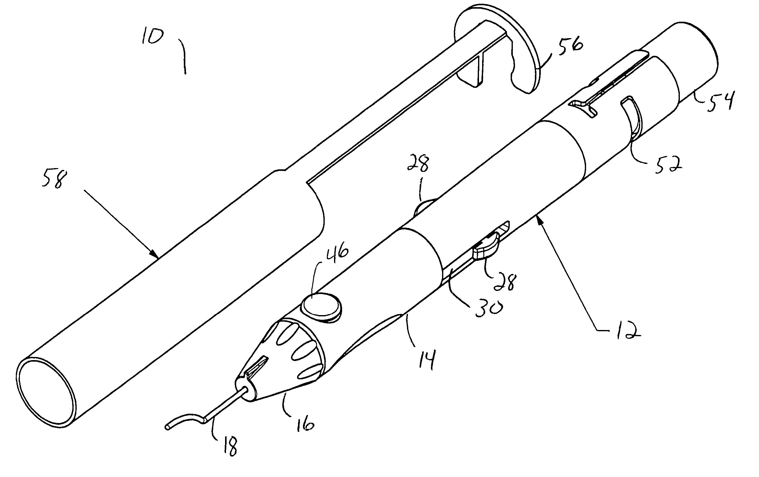
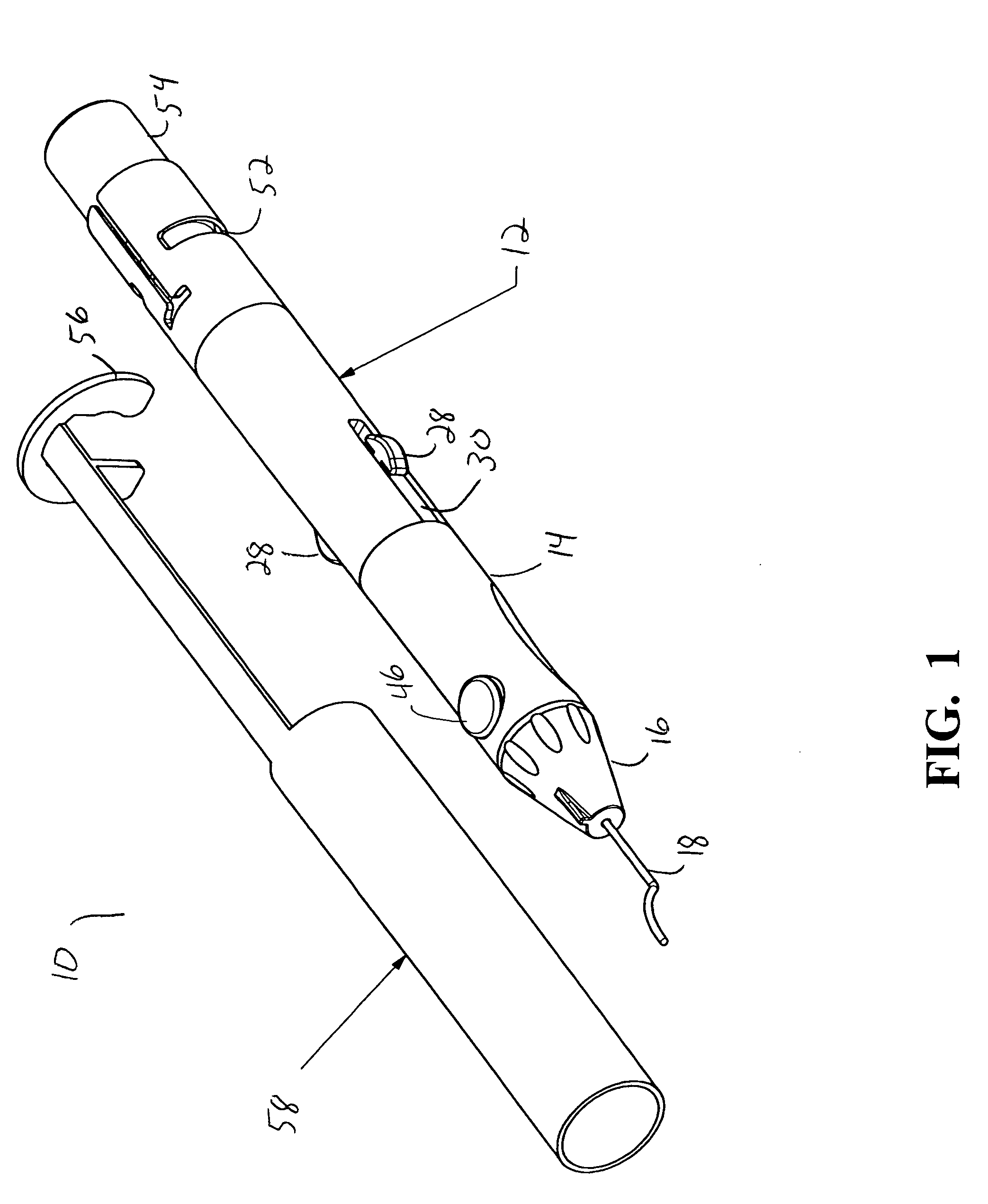
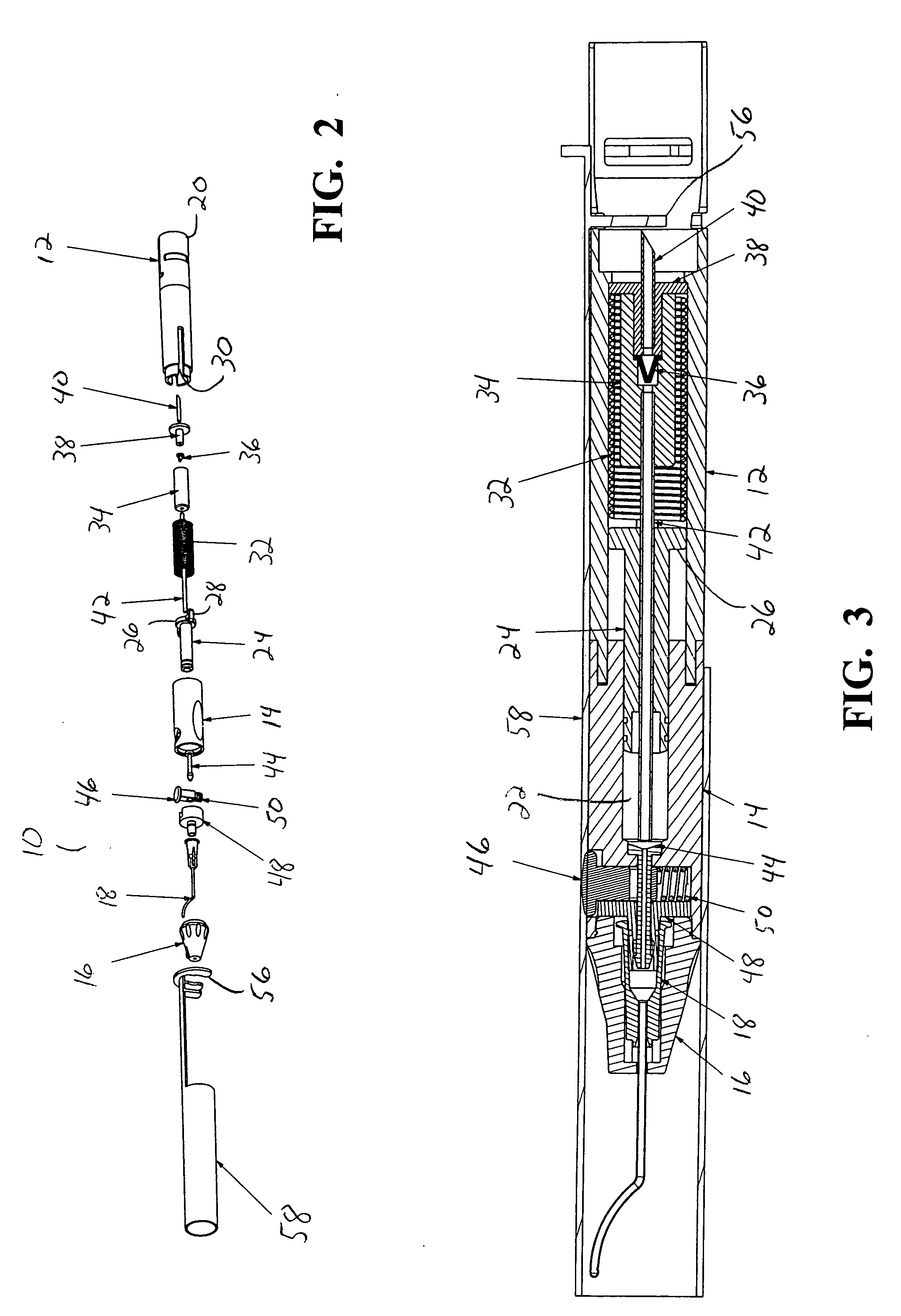
[0016] As best seen in FIGS. 1-3, drug delivery device 10 of the present invention generally comprises body 12, cylinder 14, nosecone or tip 16 and cannula 18. Body 12 is generally hollow and proximal end 20 of body 12 is generally open and sized to receive sealed vial 54 of a drug to be delivered. Cylinder 14 likewise is generally hollow having interior chamber 22. Nosecone or tip 16 mounts to cylinder 14 opposite body 12 and retains cannula 18. Cannula 18 may be any suitable cannula, such as the cannula described in U.S. Pat. No. 6,413,245 B1 (Yaacobi, et al.). Piston 24 is generally sized to reciprocate snugly within chamber 22, but contains flange 26 having a plurality of finger tabs 28 that is sized to reciprocate within body 12. Finger tabs 28 fit within slots 30 in body 12 and allow finger tabs 28 to be grasped when piston 24 is installed within device 10. Flange 26 allows piston 24 to engage spring 32, which is mounted inside body 12 between piston 24 and distal end 20. Pull...


PUM


Abstract

A syringe-like device having a chamber in which a pressurizing piston reciprocates. The chamber is connected on one end to a vial containing a drug to be delivered to an eye and on the other end to a needle or cannula for administering the drug to an eye. Pulling proximally backwards on the piston creates a vacuum in the chamber and draws the drug out of the vial. Upon release of the piston, a spring pushes the piston forward, pressurizing the chamber and forcing the drug out of the cannula. A one-way valve prevents the drug from being expelled back into the vial. A pinch valve between the chamber and the cannula allows the flow of the drug out of the cannula to be stopped. The flow rate of the drug out of the cannula can be controlled by varying the size of the chamber, piston and spring.

Description

BACKGROUND OF THE INVENTION [0001] The present invention generally pertains to the delivery of ophthalmically acceptable pharmaceutically active agents to the back of the eye and more particularly to an apparatus for sub-Tenon delivery of a drug depot to the posterior segment of a human eye proximate the macula. [0002] Several diseases and conditions of the posterior segment of the eye threaten vision. Age related macular degeneration (ARMD), choroidal neovascularization (CNV), retinopathies (e.g., diabetic retinopathy, vitreoretinopathy), retinitis (e.g., cytomegalovirus (CMV) retinitis), uveitis, macular edema, and glaucoma are several examples. [0003] Age related macular degeneration (ARMD) is the leading cause of blindness in the elderly. ARMD attacks the center of vision and blurs it, making reading, driving, and other detailed tasks difficult or impossible. About 200,000 new cases of ARMD occur each year in the United States alone. Current estimates reveal that approximately f...

Claims


Application Information

Patent Timeline
IPC IPC(8): A61M5/00
CPCA61F9/0017A61M5/1454A61M5/3213A61M2210/0612A61M2005/3115A61M2005/3128A61M5/329
Inventor HICKINGBOTHAM, DYSON W.PRESCOTT, JACK R.PRESCOTT, HEIDI A.ZICA, MICHAEL A.
Owner ALCON INC
Learn More