
Self-assembled peptide-amphiphiles & self-assembled peptide nanofiber networks presenting multiple signals

a technology of peptide nanofibers and amphiphiles, which is applied in the direction of peptides, peptide/protein ingredients, immunoglobulins, etc., can solve the problems of lack of biologically relevant signals, poor cell attachment, poor integration into the site where tissue engineered materials are utilized, etc., to promote cell-substrate adhesion, promote cell-substrate adhesion, and promote tissue repair or reconstruction

Inactive Publication Date: 2005-12-08
NORTHWESTERN UNIV
View PDF10 Cites 54 Cited by
  • Summary
  • Abstract
  • Description
  • Claims
  • Application Information

AI Technical Summary

Benefits of technology

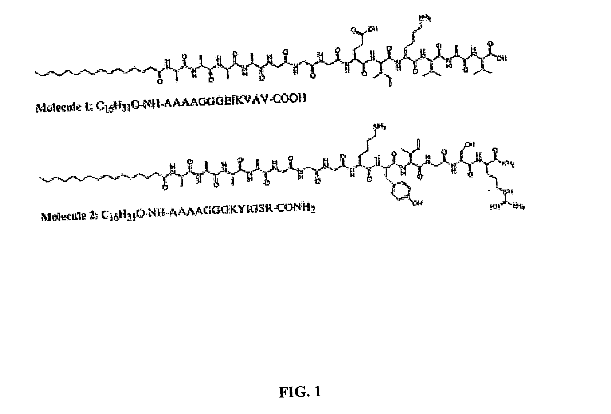
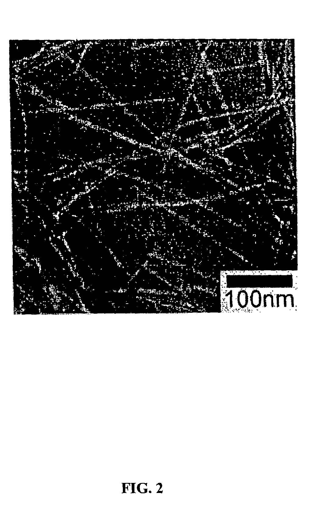
[0010] In a preferred embodiment self-assembling peptide-amphiphiles form by combining peptide-amphiphiles with sequentially different and oppositely-charged epitopes at near neutral pH, thus presenting multiple peptide signals in the same supramolecular structure. The respective peptide-amphiphile and the molecular system formed therefrom generally consist of a hydrophobic hydrocarbon tail attached to a relatively hydrophilic peptide sequence. Self-assembly of this peptide-amphiphile (PA) may be induced through pH variation (NH3, or HCl vapors), positively and negatively charged peptide amphiphiles PA+x, PA−Y where x and y are integers, divalent or polyvalent ion addition, dehydration (drying) or combinations of these among other self assembly inducing conditions. Variations of structural peptide sequences in the PA may enable the assembled nanofibers to be reversibly cross-linked for more or less structural stability, or may allow for control of the rate of self-assembly.
[0012] The versatility and functionality of this self-assembling nanofibrous material may prove to be useful in tissue repair or reconstruction. The term tissue includes muscle, nerve, vascular, and bone tissue and other common understandings of tissue. The present invention may also find application in regulation, inhibition or promotion of axon outgrowth in neurons as well as the regulation, inhibition or promotion of cell-substrate adhesion among nerve cells. The potential for coating these compositions of the present invention on surfaces, such as titanium-based orthopedic implants, may furthermore enhance existing tissue engineering strategies.

Problems solved by technology

Most polymers used today to create these scaffolds, such as polylactic acid, polyorthoesters, and polyanhydrides, are difficult to mold and, result in, among other things, poor cell attachment and poor integration into the site where the tissue engineered material is utilized.
With some exceptions, they also lack biologically relevant signals.

Method used


Image

  • Self-assembled peptide-amphiphiles & self-assembled peptide nanofiber networks presenting multiple signals
  • Self-assembled peptide-amphiphiles & self-assembled peptide nanofiber networks presenting multiple signals
  • Self-assembled peptide-amphiphiles & self-assembled peptide nanofiber networks presenting multiple signals

Examples


example 1

[0045] Materials and Methods: Abbreviations: PA: peptide-amphiphile, TEM: transmission electron microscopy, DTT: dithiothreitol, EDT: ethanedithiol, TIS: triisopropyl silane, TFA: triflouroacetic acid, HBTU: (2-(1h-benzotriazole-1-yl)-1,1,3,3-tetramethyluronium hexafluorophosphate, DiEA: Diisopropylethylamine; ESI: Electrospray ionization. Except as noted below, all chemicals were purchased from Fisher or Aldrich and used as provided. Amino acid derivatives were purchased from Applied BioSystems and NovaBiochem; derivatized resins and HBTU were also purchased from NovaBiochem. All water used was deionized with a Millipore Milli-Q water purifier operating at a resistance of 18 MW.

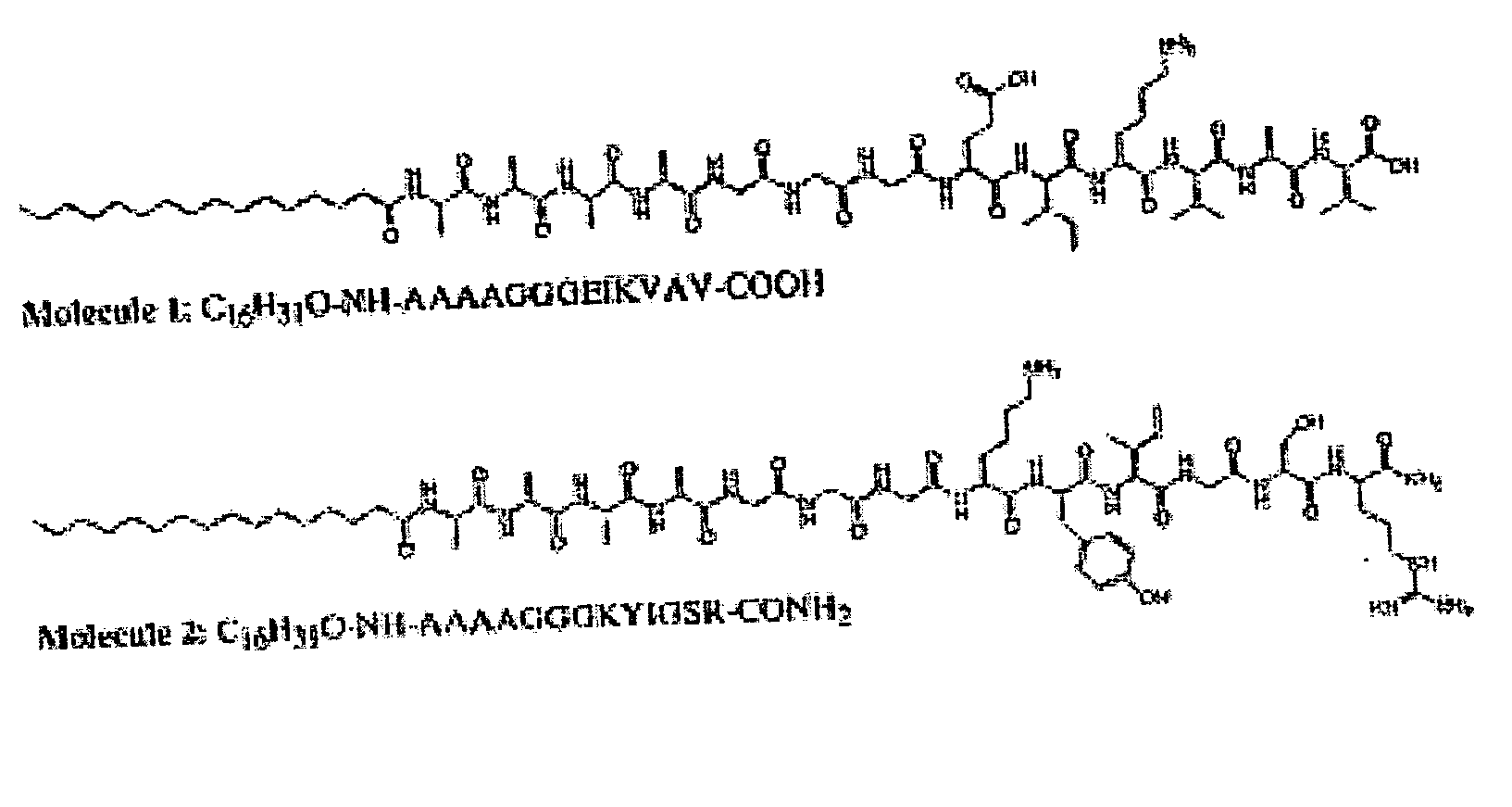
[0046] The peptide-amphiphiles were prepared on a 0.25 mmole scale using standard FMOC chemistry on an Applied Biosystems 733 A automated peptide synthesizer. Molecule 1 has a C-terminal carboxylic acid and was made using pre-derivatized Wang resin. Molecule 2 has a C-terminal amide and was made using Rink ...


PUM

PropertyMeasurementUnit
Electric chargeaaaaaaaaaa
Amphiphilicaaaaaaaaaa
Adhesion strengthaaaaaaaaaa

Abstract

The present invention provides a mixture of self-assembling peptide-amphiphiles with complementary charges whose design and function is patterned after proteins having biological functions. The oppositely charged peptide amphiphiles may be self-assembled by combining them in a charge equivalent ratio. Variations of structural peptide sequences in the oppositely charged peptide-amphiphiles enable the assembled nanofibers to exhibit two or more biologically relevant signals.

Description

CROSS-REFERENCES [0001] The present application claims priority to U.S. Provisional Patent Application Ser. No. 60 / 413,101, filed Sep. 23, 2002, the contents of which are incorporated herein by reference in their entirety.GOVERNMENT INTEREST [0002] The U.S. Government may have certain rights to this invention pursuant to Grant from the: (i) U.S. Department of Energy, Grant No. DE-FG02-00ER45810, (ii) Air Force Office of Scientific Research, Grant No. F49620-00-1-0283, and (iii) National Science Foundation, Grant No. DMR-9996253 to Northwestern University.BACKGROUND OF THE INVENTION [0003] Techniques of tissue engineering employing biocompatible scaffolds provide viable alternatives to prosthetic materials currently used in prosthetic and reconstructive surgery (e.g. craniomaxillofacial and spinal surgery). These materials also hold promise in the formation of tissue or organ equivalents to replace diseased, defective, or injured tissues. Biocompatible scaffolds can be used to form b...

Claims


Application Information

Patent Timeline
IPC IPC(8): A61K38/00A61K38/08A61K47/48C07KC07K7/06C07K7/08C07K14/78
CPCA61K9/1075A61K38/00B82Y30/00C07K7/08C07K14/78A61K47/62
Inventor STUPP, SAMUELNIECE, KRISTAHARTGERINK, JEFFREY
Owner NORTHWESTERN UNIV
Learn More