Marker for stem cells and its use
- Summary
- Abstract
- Description
- Claims
- Application Information
AI Technical Summary
Benefits of technology
Problems solved by technology
Method used
Image
Examples
example 1
Detection of integrin alpha10 and integrin alpha11 chain on human MSC
Objective
[0182] The objective of this example is to analyse human MSC for the expression of integrin alpha10 and alpha11, using immunoprecipitation.
Materials and Methods
[0183] Human mesenchymal stem cells (obtained from In Vitro, Sweden, at passage 2), were cultured in MSCBM medium (provided by In Vitro, Sweden) until passage 4 and then surface biotinylated.
[0184] In brief, cells adherent on the plate were washed once with PBS and then surface biotinylated using 0.5 mg / ml Sulfo-NHS-LC-biotin (Pierce) in 4 ml PBS for 20 min. Cells were then washed once with PBS and 10 ml 0.1 M glycine / PBS were added for 5 min.
[0185] After washing once with PBS cells were lysed in 1 ml lysis buffer (1% NP40, 10% glycerol, 20 mM Tris / HCl, 150 mM NaCl, 1 mM MgCl2, 1 mM CaCl2, protease inhibitor cocktail BM, pH7.5). The cell lysate was collected with a plastic scraper, pipetted into an eppendorf tube and spun down for 10 min at 1...
example 2
Identification of HEK293 cells expressing the integrin alpha10beta1 and the alpha11beta1.
Objective
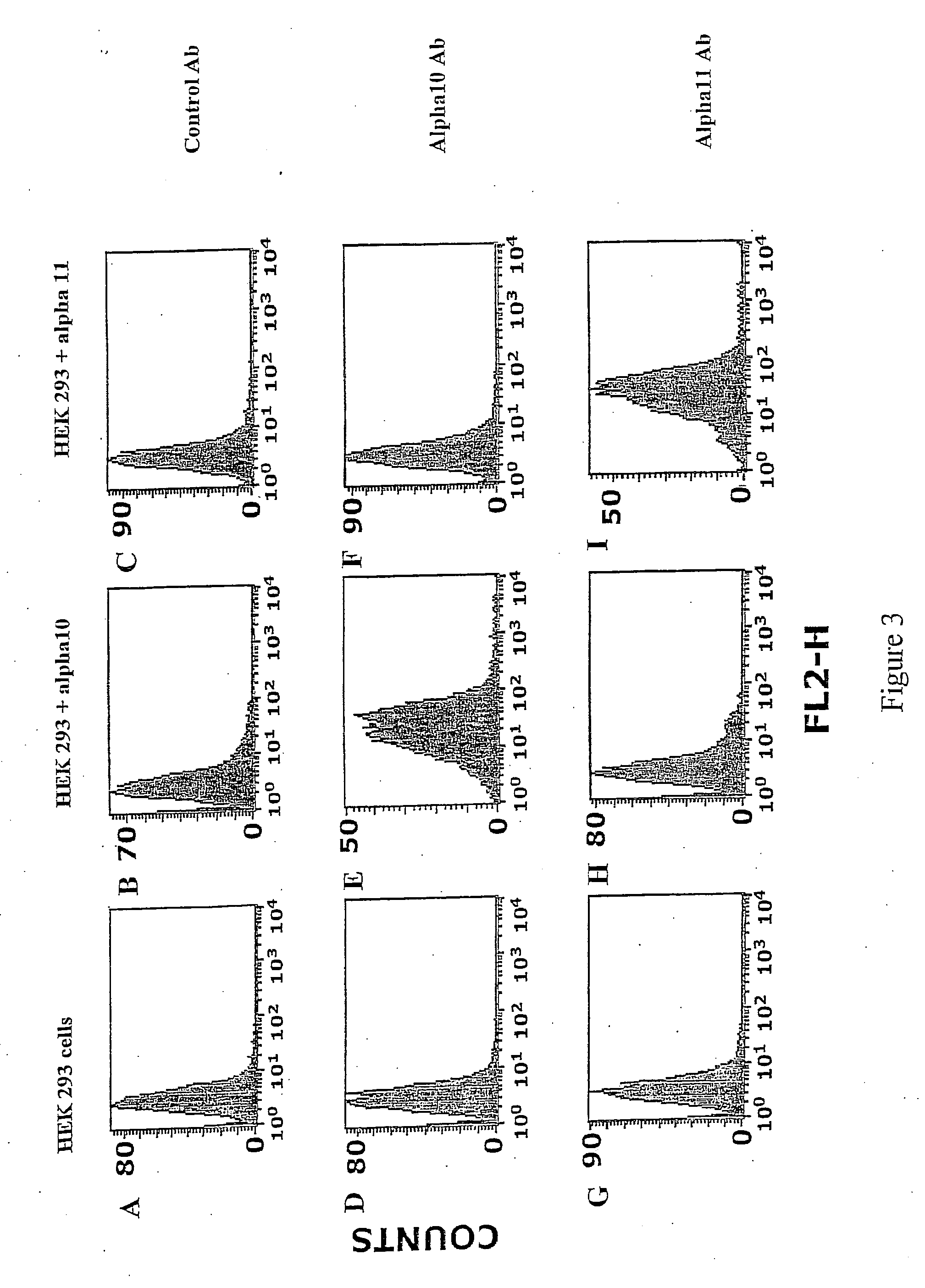
[0195] The objective with this example is to use antibodies to alpha11 and alpha11 to identify and differentiate between HEK293 cells expressing the integrin alpha10beta1 and the integrin alpha11beta1.
Materials and Methods
[0196] Integrin alpha10 and alpha11-transfected HEK293 cells and non-transfected HEK293 cells were trypsinized, washed with PBS and then incubated for 20 min with integrin antibodies against alpha10 and alpha11 (1 μg / ml in PBS supplemented with 1%BSA).
[0197] Labelled cells were washed twice with PBS / 1%BSA and then incubated for 20 min with PE labelled goat-anti-mouse Ig (Phrmingen, BD Biosciences) at a concentration of 1g / ml in PBS / 1%BSA.
[0198] Cells were thereafter washed twice in PBS / 1%BSA and were analysed on a FACSort® (Becton-Dicldnson) by collecting 10,000 events with the Cell Quest® software program (Becton-Dickinson).
Results
[0199] The results are show...
example 3
Identification of MSC expressing the integrin alpha10 from human colony forming cells derived from human bone marrow.
Objective
[0205] To test whether colony-forming cells derived from human bone marrow express the integrin alpha10 and represent a population of mesenchymal stem cells.
Materials and Methods
[0206] Human mononuclear bone marrow cells were isolated from bone marrow by density centrifuigation.
[0207] 30×106 cells were taken in 20 ml medium (MSCGM medium provided by Poietics and delivered via lnvitro, comprising 440 ml MSCBM (lot 017190) and 2×25ml MCGS (lot 092295) and L-glutamine and Penicillin / Streptomycin) into a T75 flask and incubated in the cell incubator.
[0208] Cells were grown until day 12 (medium was changed twice) and then trypsinized and split (5000 cells / cm2). In general, cells were split at 90% confluency.
[0209] Cells were grown for a further 3 days and then split again (5000 cells / cm2). At this point the influence of FGF-2 on alpha10-expression on hMSC ...
PUM
| Property | Measurement | Unit |
|---|---|---|
| Length | aaaaa | aaaaa |
| Length | aaaaa | aaaaa |
| Length | aaaaa | aaaaa |
Abstract
Description
Claims
Application Information
 - R&D Engineer
- R&D Manager
- IP Professional
- Industry Leading Data Capabilities
- Powerful AI technology
- Patent DNA Extraction
Browse by: Latest US Patents, China's latest patents, Technical Efficacy Thesaurus, Application Domain, Technology Topic, Popular Technical Reports.
© 2024 PatSnap. All rights reserved.Legal|Privacy policy|Modern Slavery Act Transparency Statement|Sitemap|About US| Contact US: help@patsnap.com


