Method of inhibiting choroidal neovascularization
a choroidal neovascularization and choroidal neovascularization technology, applied in the direction of drug compositions, antiparasitic agents, extracellular fluid disorder, etc., can solve problems such as vision loss and possibly blindness, and achieve the effect of reducing the loss of visual acuity and rapid identification of additional anti-angiogenic agents
- Summary
- Abstract
- Description
- Claims
- Application Information
AI Technical Summary
Benefits of technology
Problems solved by technology
Method used
Image
Examples
example 1
Matrigel™ Based Animal Model
[0042] Within this application, unless otherwise stated, the techniques utilized may be found in any of several well-known references, such as: Molecular Cloning: A Laboratory Manual (Sambrook et al., Cold Spring Harbor Laboratory Press (1989); “Guide to Protein Purification” in Methods in Enmology (M. P. Deutshcer, ed., (1990) Academic Press, Inc.); Culture of Animal Cells: A Manual of Basic Technique, 2nd Ed., Liss, Inc., New York, N.Y., (1987).
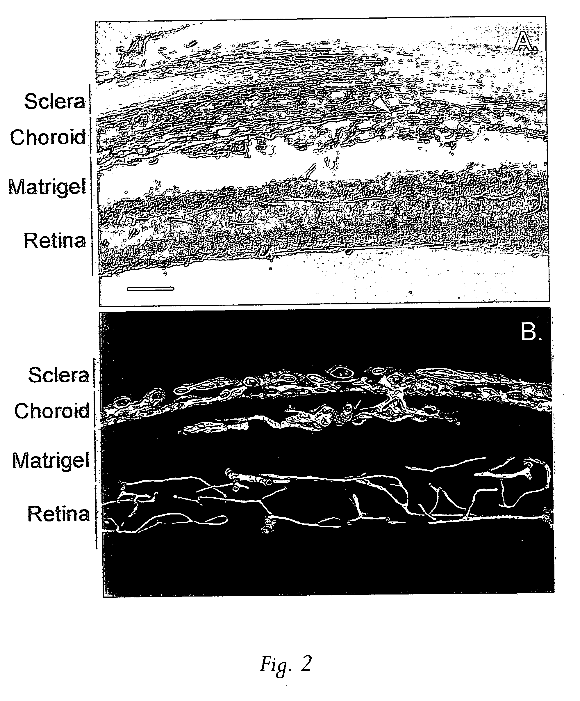
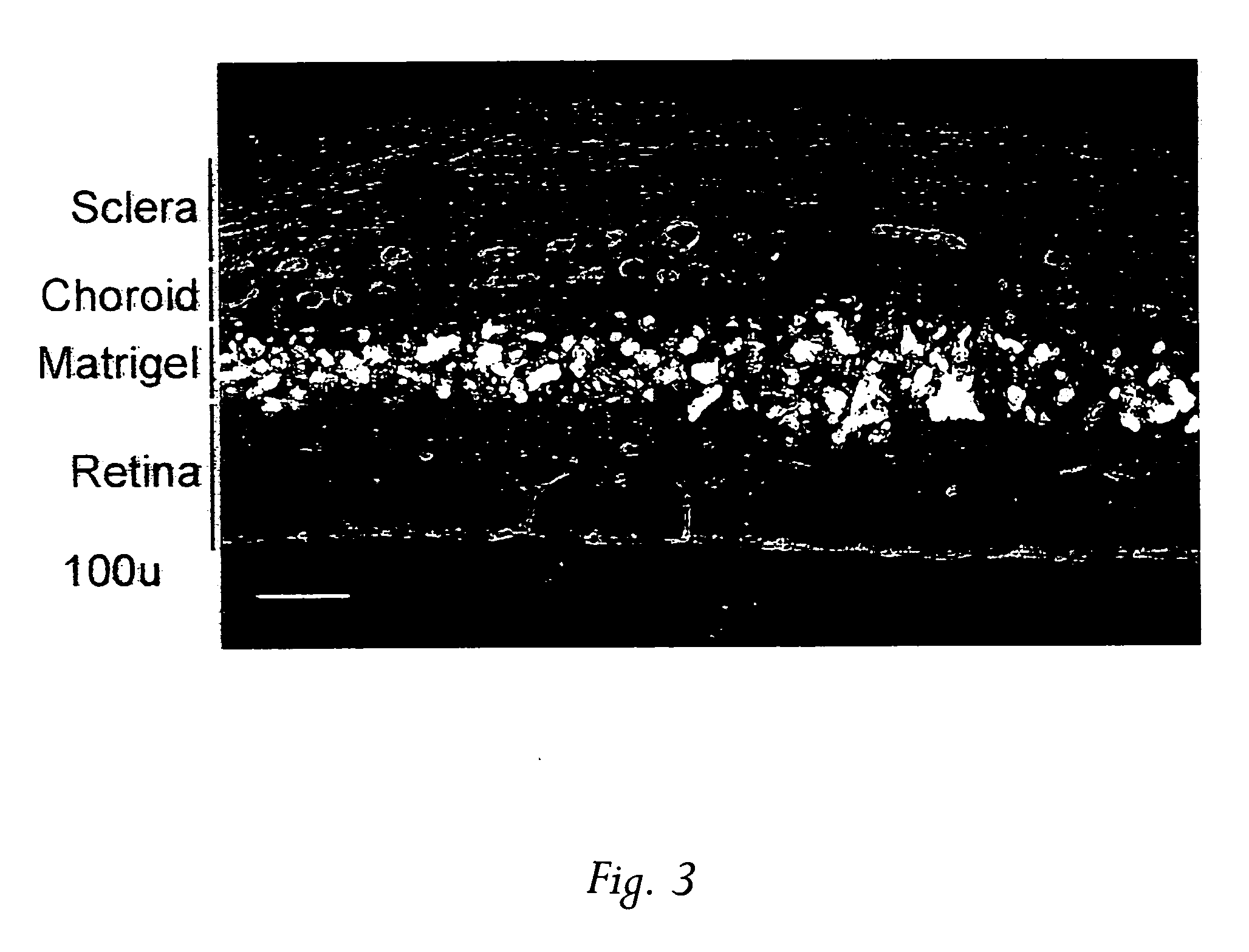
[0043] In search of effective treatments for CNV, a simple animal model was created by injecting 2-3 μl Matrigel™ to the subretinal space of adult Sprague-Dawley rats using a 33-guage needle connected to a 10 μl Hamilton microsyringe. A week or more later the animals were sacrificed by CO2 inhalation and perfused with Vessel Paint, a new visualization technique recently developed in the inventors' lab that enables screening agents, including chemical compounds and proteins, for their potential in inhibiting CNV...
example 2
Inhibition of CNV
[0047] In the initial characterization of the model, it was suspected that there was a possible involvement of an inflammatory reaction to Matrigel™ in the generation of neovascularization. As a result, two known immunosuppressants were tested, cyclosporin and raparnycin.
[0048] Oral administration of cyclosporin (15 mg / kg / d, given 4 day before Matrigel™ injection thorough 10 days after injection) had no effect on CNV. In marked contrast, however, oral rapamycin (Rapamune®, 1.5 mg / kg / d, given 4 days before Matrigel™ injection thorough 10 day after injection) resulted in complete inhibition of CNV development in the 16 eyes tested. Rapamycin is commercially available as oral solution, marketed as Rapamune Oral Solution by Wyeth-Ayerst. Thus, the anti-CNV properties of rapamycin by local administration were further examined.
[0049] Since rapamycin is not soluble in water, it was either dissolved in DMSO (tested in 8 eyes) or suspended in PBS (tested in 6 eyes), then ...
example 3
Vessel Painting
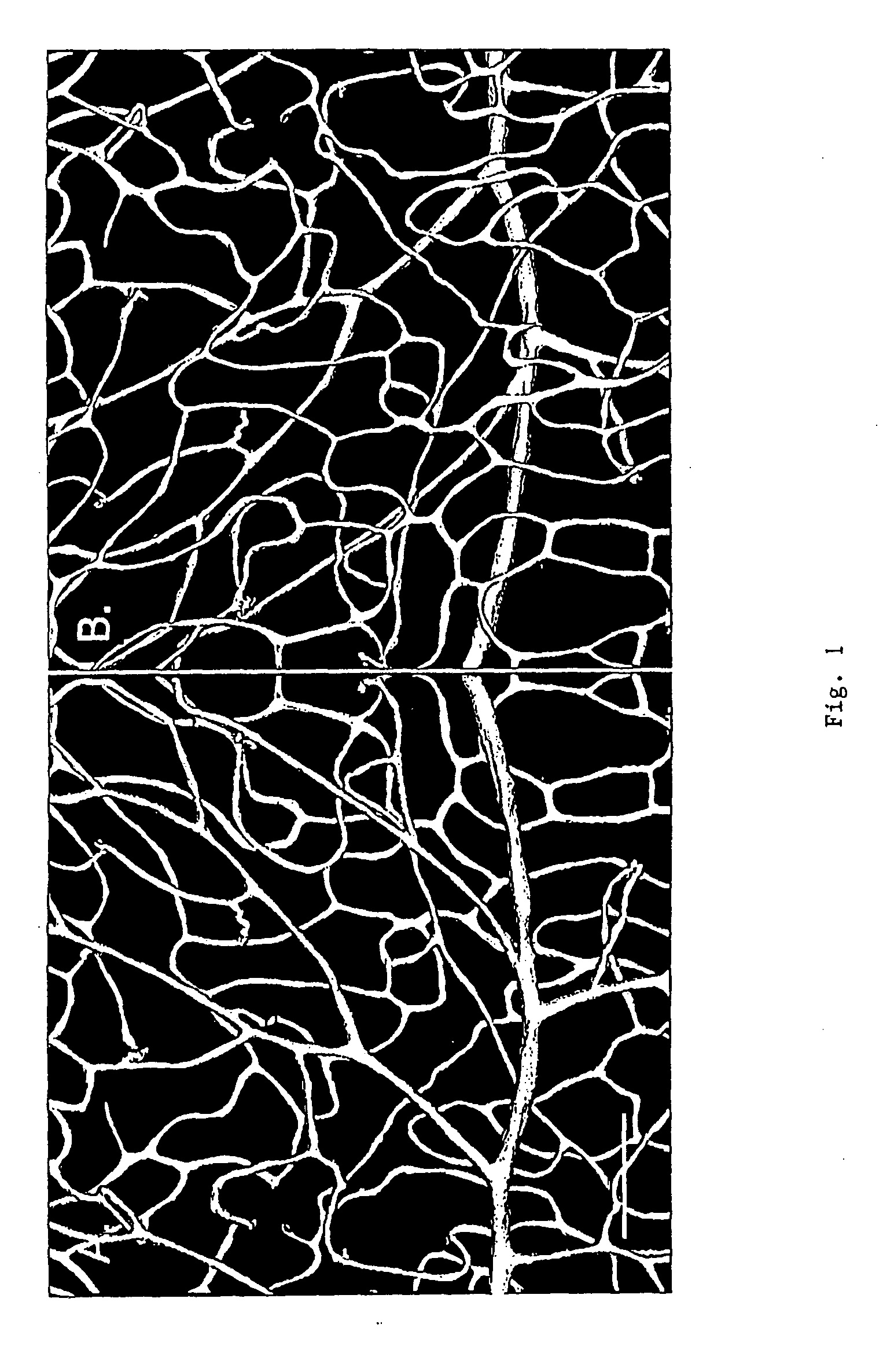
[0052] The retina from a normal 3 months old Sprague-Dawley rat, sacrificed by CO2 overdose was perfused with Vessel Paint (DiI, 0.1 mg / ml), followed by 4% paraformaldehyde. The retina was dissected and postfixed in the same fixative for 1 hr, rinsed in phosphate buffered saline (PBS), and flat-mounted on glass slides. Microphotographs were taken on a Nikon E800 microscope.
[0053] Microphotographs of the flat-mount retina showed that vessels stain brightly with a low background. Endothelial cell nuclei were also stained and easily identifiable. The spatial structure of the vasculature was well preserved and the deep retinal capillaries were also visible.
[0054] The vascular network is better appreciated by images to show its three-dimensionality. Excellent 3-D images of vasculature can be obtained using the corrosion-casting technique and scanning electron microscopy (SEM), see Konerding M A (1991) Scanning electron microscopy of corrosion casting in medicine. Scanni...
PUM
| Property | Measurement | Unit |
|---|---|---|
| pH | aaaaa | aaaaa |
| thick | aaaaa | aaaaa |
| angle | aaaaa | aaaaa |
Abstract
Description
Claims
Application Information
 - R&D
- Intellectual Property
- Life Sciences
- Materials
- Tech Scout
- Unparalleled Data Quality
- Higher Quality Content
- 60% Fewer Hallucinations
Browse by: Latest US Patents, China's latest patents, Technical Efficacy Thesaurus, Application Domain, Technology Topic, Popular Technical Reports.
© 2025 PatSnap. All rights reserved.Legal|Privacy policy|Modern Slavery Act Transparency Statement|Sitemap|About US| Contact US: help@patsnap.com


