Classification of breast lesion method and system

a breast lesion and classification technology, applied in the field of imaging systems, can solve the problems of inability to automate the implementation of tools and techniques in image processing to extract these features and quantify them for accurate classification, and the inability to achieve accurate classification, so as to and enhance the quality of the image

Inactive Publication Date: 2005-06-16
GENERAL ELECTRIC CO
View PDF12 Cites 21 Cited by
  • Summary
  • Abstract
  • Description
  • Claims
  • Application Information

AI Technical Summary

Benefits of technology

[0007] Briefly, in accordance with one aspect of the present invention, an automated technique for determining a plurality of characteristics of a breast lesion is provided. The technique comprises automatically identifying a region of interest in an image, the region of interest comprising the breast lesion. The technique further comprises preprocessing the region of interest to enhance a quality of the image, and automatically segmenting the breast lesion in the region of interest. The technique further comprises automatically measuring a plurality of measurements for determining the plurality of characteristics of the breast lesion, and automatically classifying the breast lesion as benign or malignant based on the plurality of measurements.
[0008] In a further embodiment, a system for determining a plurality of characteristics of a breast lesion is provided. The system comprises a memory unit configured for storing an image and a processor configured for automatically identifying a region of interest in the image, the region of interest comprising the breast lesion. The processor is further configured for preprocessing the region of

Problems solved by technology

Satisfactory techniques for such discrimination are not, however, yet available.
Usually, solid breast masses are hard to distinguish from malignant masses through traditional image-based diagnosis techniques.
However, the tools and techniques in image processing to extract these features and quantify them for accurate classification are not available for automated implementation, as the tools are manual or sem

Method used


Image

  • Classification of breast lesion method and system
  • Classification of breast lesion method and system
  • Classification of breast lesion method and system

Examples


Embodiment Construction

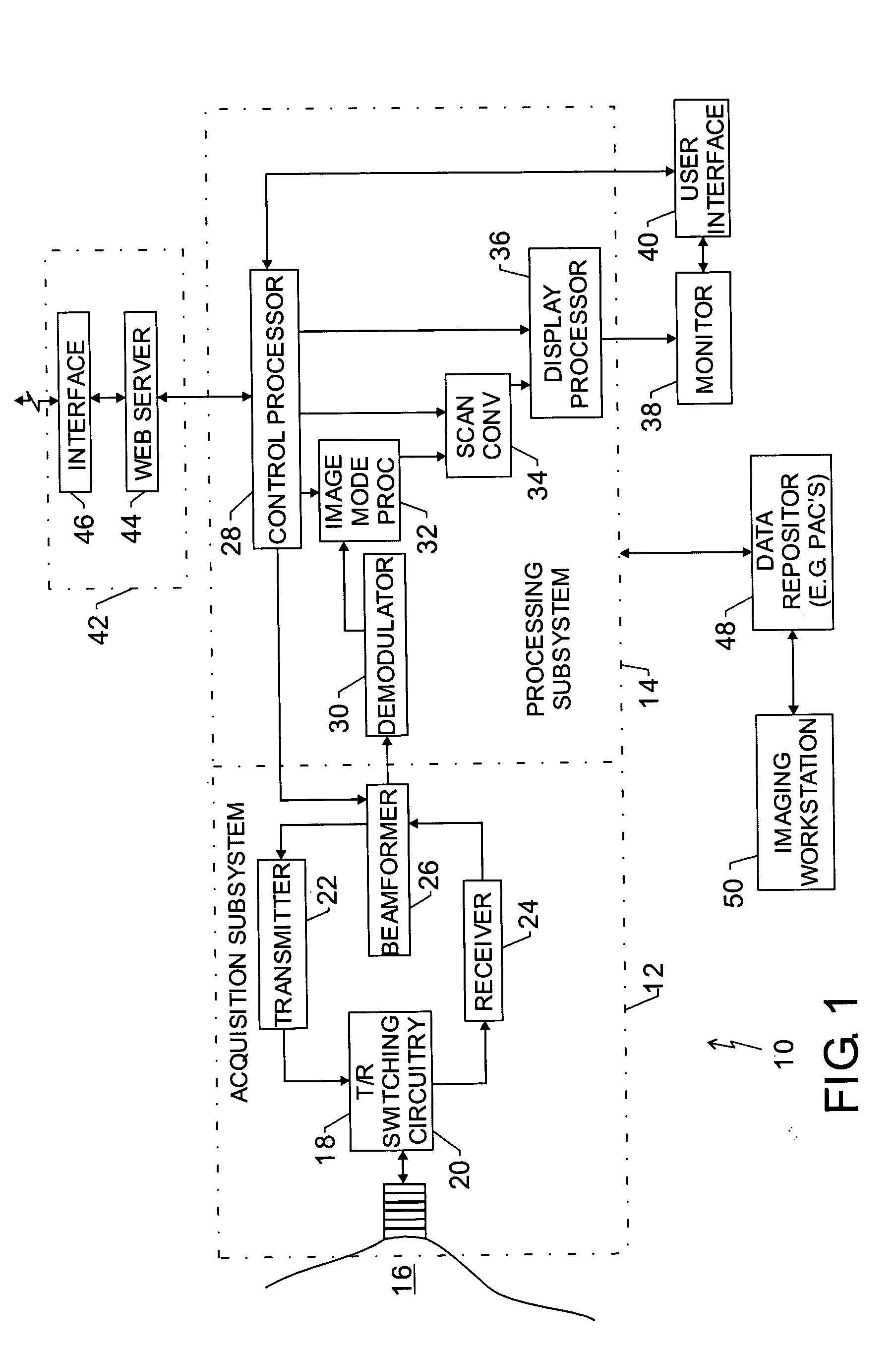
[0018]FIG. 1 is a block diagram of an embodiment of an ultrasound system 10 implemented in accordance to one aspect of the invention. The ultrasound system comprises of acquisition subsystem 12 and processing subsystem 14. The acquisition subsystem 12 comprises a transducer array 18 (comprising a plurality of transducer array elements), transmit / receive switching circuitry 20, a transmitter 22, a receiver 24, and a beamformer 26. Processing subsystem 14 comprises a control processor 28, a demodulator 30, an imaging mode processor 32, a scan converter 34 and a display processor 36. The display processor is further coupled to a monitor for displaying images. User interface 40 interacts with the control processor and the display monitor. The control processor may also be coupled to a remote connectivity subsystem 42 comprising a web server 44 and a remote connectivity interface 46. Processing subsystem may be further coupled to data repository 48 to receive ultrasound image data. The d...


PUM


Abstract

An automated method for determining a plurality of characteristics of a breast lesion is provided. The method comprises automatically identifying a region of interest in an image, the region of interest comprising the breast lesion, and preprocessing the region of interest to enhance a quality of the image. The method further comprises automatically segmenting the breast lesion in the region of interest and automatically measuring a plurality of measurements for determining the plurality of characteristics of the breast lesion. The breast lesion is automatically classified as benign or malignant based on the plurality of measurements.

Description

BACKGROUND OF THE INVENTION [0001] The invention relates to imaging systems and more specifically to a method and apparatus for classification of breast lesions. [0002] Breast cancer is one of the leading causes of death among women. Typically, breast cancer is detected by a method called mammography, generally an X-ray imaging procedure. Mammography has become the standard for detecting and characterizing malignancy. Following image acquisition, a radiologist typically reads the images to discern whether suspicious growths appear, and whether they are likely problematical. However, in recent times ultrasound imaging techniques has shown promise for discriminating cysts from solid masses. Satisfactory techniques for such discrimination are not, however, yet available. [0003] Usually, solid breast masses are hard to distinguish from malignant masses through traditional image-based diagnosis techniques. As a result, patients in which such masses are detected are usually referred for b...

Claims


Application Information

Patent Timeline
IPC IPC(8): A61B5/107A61B8/08G06K9/00G06T7/00
CPCA61B5/1075A61B5/7264G06T2207/30068G06T7/0012A61B8/0825G16H50/20
Inventor KAMATH, VIDYA PUNDALIKTHOMENIUS, KAI ERIKBAKTHAVATHSALU, MANNAN
Owner GENERAL ELECTRIC CO
Why Eureka
Try Eureka