
Multiple biopsy device

a biopsy device and multi-functional technology, applied in the field of medical devices, can solve the problems of loss of traceability, added wear and tear on the endoscope, and time-consuming process

Inactive Publication Date: 2005-06-09
BOSTON SCI SCIMED INC
View PDF5 Cites 19 Cited by
  • Summary
  • Abstract
  • Description
  • Claims
  • Application Information

AI Technical Summary

Benefits of technology

The patent describes a device that can take multiple tissue samples from a patient using an endoscope. The device has a cap with multiple chambers where the tissue samples are collected. A cutter is used to cut the tissue that enters a chamber, and then the chamber is sealed and another chamber is positioned to collect another tissue sample. The technical effect of this device is that it allows for more efficient and accurate collection of tissue samples from a patient.

Problems solved by technology

This process is not only time consuming but adds wear and tear on the endoscope caused by the repeated insertion and withdrawal of the forceps.
However, they are not commonly used because the samples may commingle resulting in a loss of traceability.
In addition, there is some chance that the samples may cross contaminate each other.

Method used


Image

  • Multiple biopsy device
  • Multiple biopsy device
  • Multiple biopsy device

Examples


Embodiment Construction

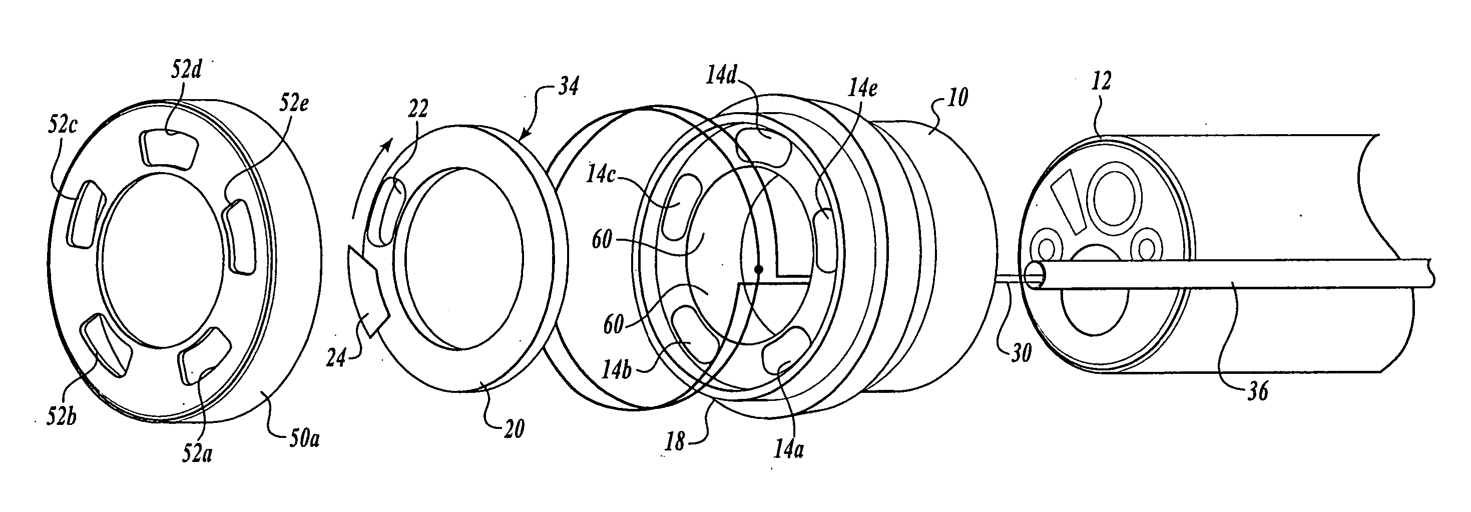
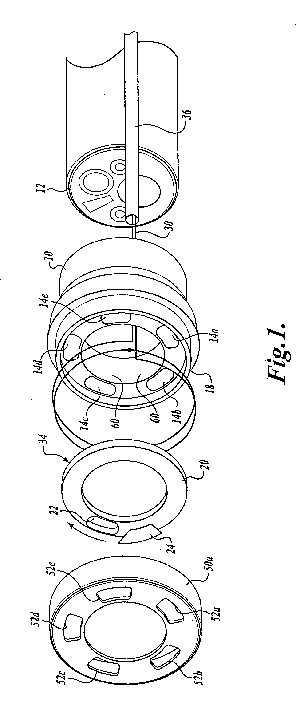
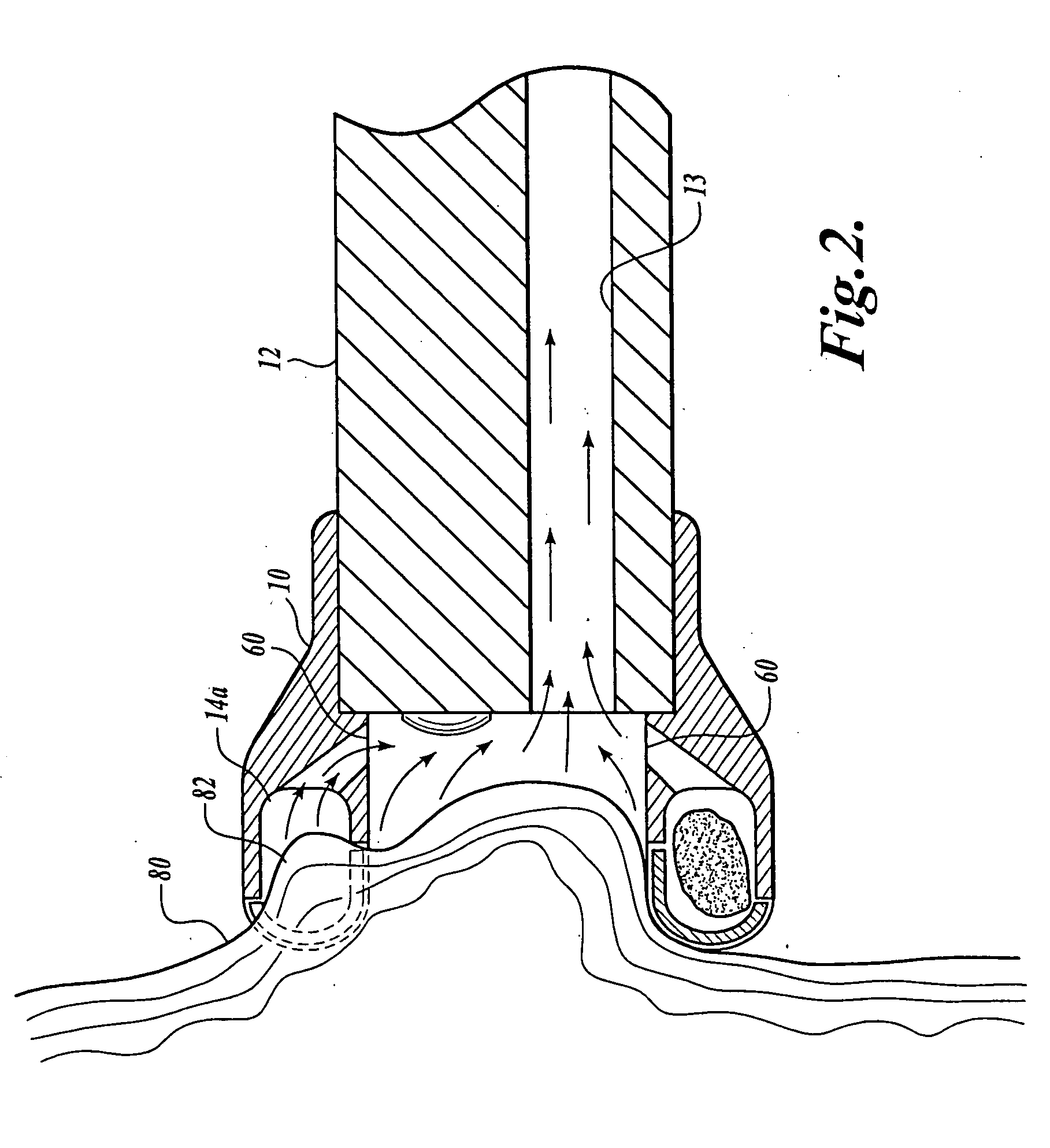
[0011] In accordance with one embodiment of the present invention, a multiple biopsy device 10 comprises a cap that is fitted over the distal end of a conventional endoscope / bronchoscope 12 or other type of device that lets a physical visually examine internal body cavities. The biopsy device 10 has a lumen passing through the center with two different radiuses. At the proximal end, the lumen is sized such that it will snugly fit over the outer diameter of the endoscope. The lumen has a second, narrower, diameter toward the distal end that forms a step that engages the end of the endoscope such that the biopsy device cannot slide along the length of the endoscope. Preferably, the biopsy device 10 is made of a polymer or other biocompatible material and is secured to the distal end of the endoscope 12 with a friction fit.

[0012] The biopsy device 10 includes a number of chambers 14A, 14B, 14C . . . 14E disposed about its periphery. Each chamber has an opening that is oriented in the ...


PUM


Abstract

A multiple biopsy device has a number of chambers into which tissue samples are received. A cutting mechanism, such as a blade, moves relative to the chambers to cut a tissue sample that is within a chamber. In one embodiment of the invention, the multiple biopsy device is removably secured to the distal end of an endoscope / bronchoscope.

Description

CROSS-REFERENCE TO RELATED APPLICATION [0001] The present application is a continuation of U.S. patent application Ser. No. 10 / 118,202, filed Apr. 5, 2002, the benefit of which are claimed under 35 U.S.C. § 120 and are herein incorporated by reference.FIELD OF THE INVENTION [0002] The present invention relates to medical devices and in particular to devices for obtaining biopsy samples from interior body cavities. BACKGROUND OF THE INVENTION [0003] In addition to obtaining a visual inspection with an endoscope / bronchoscope, etc., many physicians will biopsy a region of interest in order to confirm or deny the presence of disease. [0004] The conventional method of obtaining a tissue sample is with a biopsy forceps. Most forceps have an elongated shaft that terminates at a sharp cutter with a tissue receptacle at the distal end. The forceps are threaded through a biopsy channel of the endoscope and a tissue sample is obtained. The forceps are then withdrawn from the endoscope and an a...

Claims


Application Information

Patent Timeline
Patent Type & Authority Applications(United States)
IPC IPC(8): A61B10/00A61B10/02A61B17/30A61B17/32A61B19/00
CPCA61B10/0266A61B10/0275A61B10/0283A61B2019/444A61B2017/00296A61B2017/306A61B2017/32004A61B2010/0225A61B90/92
Inventor DAMARATI, JOHN JAIRO
Owner BOSTON SCI SCIMED INC
Learn More