PTD-modified proteins
a technology of modified proteins and proteins, applied in the field of ptdmodified proteins, can solve the problems of inability to treat inherited genetic diseases of the brain, inability to approach global therapy for degenerative diseases due to polyglutamine repeat expansion or channel mutation, etc., and achieve the effect of gene replacement therapy
- Summary
- Abstract
- Description
- Claims
- Application Information
AI Technical Summary
Benefits of technology
Problems solved by technology
Method used
Image
Examples
example 1
Production of Recombinant vectors
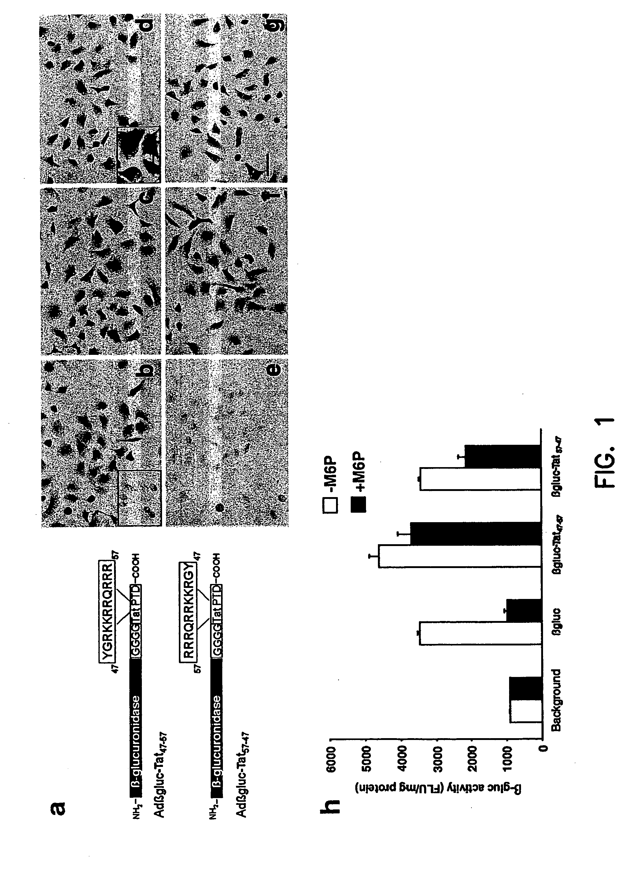
[0092] Primer 1 (5′-AAACTCGAGATGGCCCGGGGGTCGGCGGTTGCC-3′) (SEQ ID NO: 1) and primer 2 (5′-TGCTCTAGATCATCTTCGTCGCTGTCTCCGCTTCTTCCTGCCATAACCGCC ACCG-CCAGTAAACGGGCTGTT T TCCAAACA-3′) (SEQ ID NO:2) were used to create the β-glucuronidase-Tat47-57 fusion protein. Primer 1 and primer 3 (5′TGCTCTAGATCAATAGCCCCTCTTC TTCCGTCT CTGTCGTCGTCTACCGCCACCGCCAGTAAACGGGCTGTTTTCCA AACA-3′) (SEQ ID NO:3) were used to make the β-glucuronidase-Tat57-47 fusion protein. PCR fragments were digested with XhoI and XbaI and the fragments cloned into similarly cut E1 shuttle plasmids (pPacRSVKpnA; described in (Anderson, et al., (2000) Gene Ther. 7(12):1034-1038)). The resultant plasmids were named pPacRSVβGluc-Tat PTD47-57 or pPacRSV βGluc-Tat PTD57-47. Adenoviruses with β-glucuronidase, β-glucuronidase-Tat PTD47-57 or β-glucuroniase-Tat PTD57-47 in E1 and eGFP in E3 were produced by co-transfecting PacI linearized pPacRSVβGluc-Tat PTD47-57, pPacRSVβGluc-Tat PTD57-47 or pPacRSV...
example 2
In vitro Studies
[0093] HeLa cells were infected with Adβgluc-Tat47-57, Adβgluc-Tat57-47 or control Adβgluc at 20 infectious units (i.u.) / cell and supernatants harvested 72 h later. β-Glucuronidase activity was quantified using the previously described fluorometric assay. Briefly, aliquots were reacted in 10 mM 4-methylumbellifryl-β-D-glucuronidase (Sigma, St. Louis, Mo.) in 0.1 M sodium acetate (pH 4.8) for 1 h at 37° C. Reactions were stopped by addition of 2 ml of 320 mM glycine in 200 mM carbonate buffer, pH 10.0 (Glaser, et al., (1973) J.Lab.Clin.Med. 82:969-977).
[0094] Fluorescence was measured at 415 mn after excitation at 360 nm (TD-700 Fluorometer; Turner Design, Sunnyvale, Calif.). β-Glucuronidase activity is expressed as nanomoles of 4-methylumbellferone released per hour (FLU) per mg protein. Purified β-glucuronidase (kindly provided by William Sly, Washington University, St. Louis Mo.) was used as standard. Protein concentrations were determined using the Bio-Rad DC pr...
example 3
In vivo Studies
[0096]β-Glucuronidase-deficient mice were obtained from the Jackson Laboratory 30 (Bar Harbor, Me.) and from our own breeding colony. The genotype for the latter was confirmed by morphological and genetic analyses. The animals were between 8 and 10 weeks old and weighed 16-24 g. C57BL / 6 wild-type mice were purchased from Harlan Sprague (Indianapolis, Ind.).
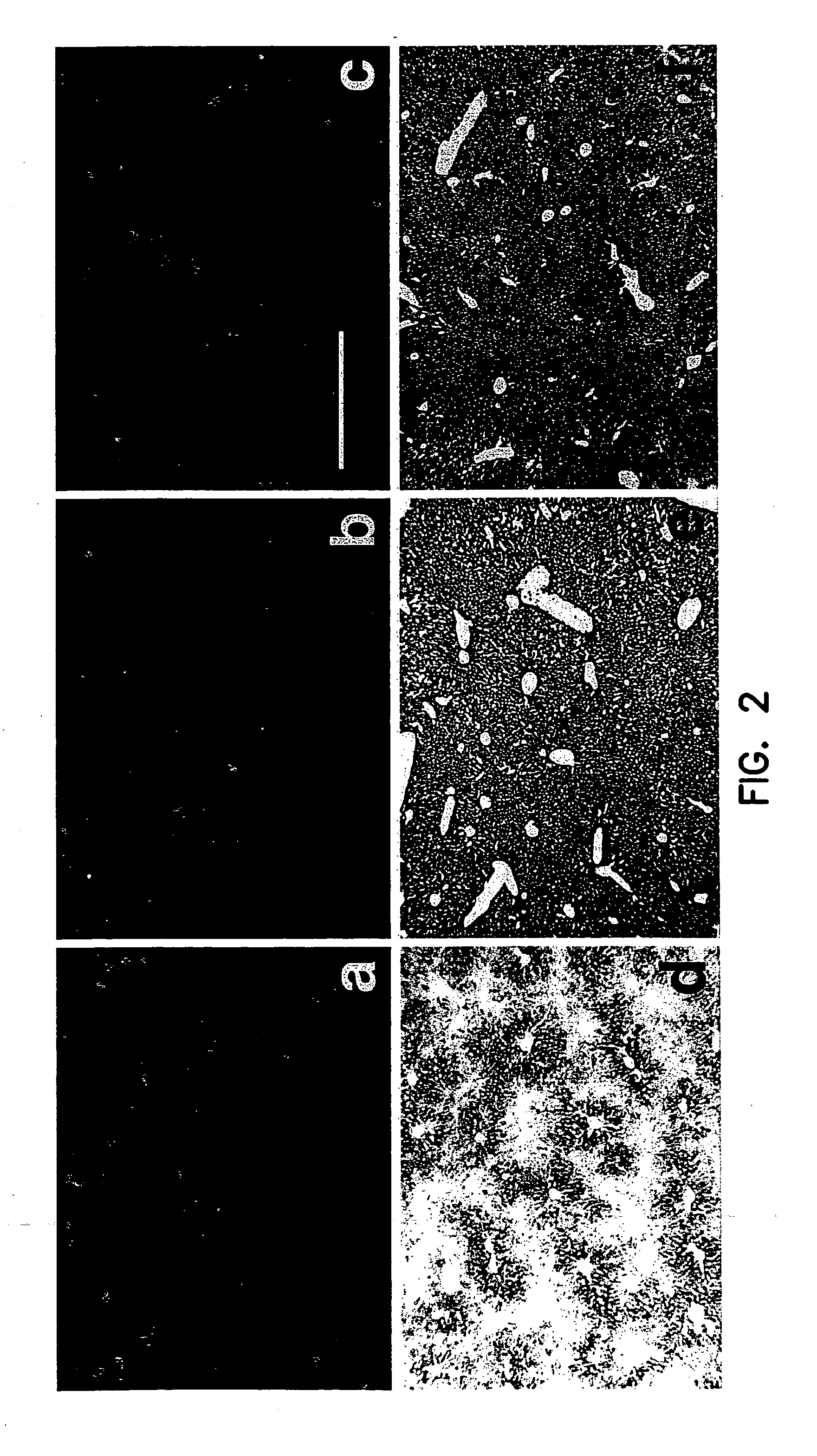
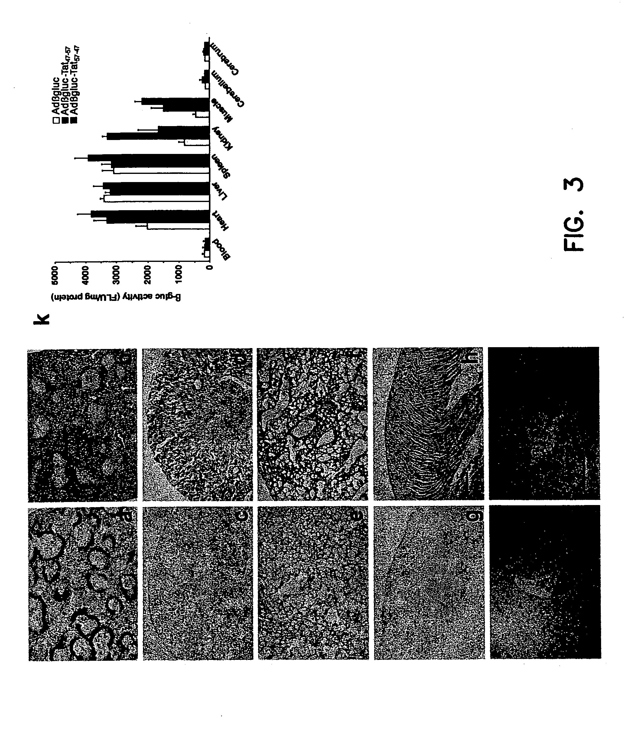
[0097] Adβgluc-Tat47-57, Adβgluc-Tat57-47 or Adβgluc were injected into the tail vein (2×10⁹ i.u.) of β-glucuronidase deficient mice. Adβgluc-Tat47-57, Adβgluc-Tat57-47 or Adβgluc (2×10⁷ i.u. total) were injected into the right striatum or right lateral ventricle of C57BL / 6 mice or β-glucuronidase deficient mice as described earlier (Stein, et al., (1999) J. Virol. 73(4):3424-3429). Animals were sacrificed 10 days after intravenous (n=3 / group), striatal (n=5 / group) or ventricular injection (n=5 / group). Tissues were sonicated, placed in lysis solution (Sands, et al., (1994) J.Clin.Invest. 93:2324-2331) and centrifu...
PUM
| Property | Measurement | Unit |
|---|---|---|
| Solubility (mass) | aaaaa | aaaaa |
Abstract
Description
Claims
Application Information
 - R&D
- Intellectual Property
- Life Sciences
- Materials
- Tech Scout
- Unparalleled Data Quality
- Higher Quality Content
- 60% Fewer Hallucinations
Browse by: Latest US Patents, China's latest patents, Technical Efficacy Thesaurus, Application Domain, Technology Topic, Popular Technical Reports.
© 2025 PatSnap. All rights reserved.Legal|Privacy policy|Modern Slavery Act Transparency Statement|Sitemap|About US| Contact US: help@patsnap.com


