
Suppression of non-biological motion

Inactive Publication Date: 2005-05-19
BAHNSON ALFRED BLALOCK
View PDF0 Cites 1 Cited by
  • Summary
  • Abstract
  • Description
  • Claims
  • Application Information

AI Technical Summary

Benefits of technology

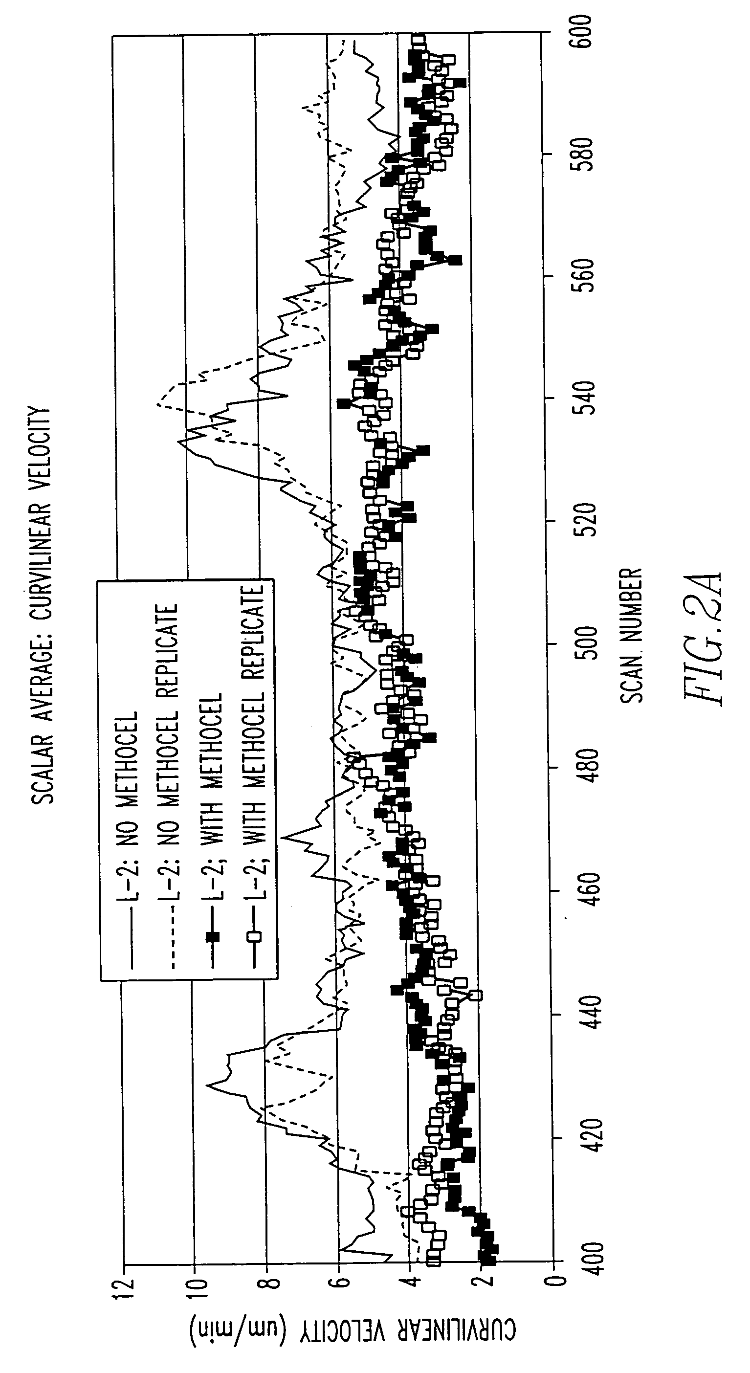
[0038] The present invention pertains to a method for analyzing a cell by suppressing non-biological movement. The method comprises the steps of placing the cell in a solution. There is the step of using methyl cellulose in the solution for reducing or eliminating the effects of micro-turbulances due to thermal convection in the solution.
[0040] The present invention pertains to a method for analyzing a cell. The method comprises the steps of placing the cell in a solution. There is the step of introducing a viscous fluid having a viscosity of about 100-5000 centipose in the solution for stopping or reducing the effects of gravity on the cell.
[0041] The present invention pertains to a method for analyzing a cell. The method comprises the steps of placing the cell in a solution. There is the step of introducing a viscous fluid having a viscosity of about 100-5000 centipose in the solution for reducing the effects of micro-turbulences due to thermal convection.

Problems solved by technology

Of particular interest to us is the possibility of screening for very short-lived secreted products on the basis of changes in migration patterns or morphology or phenotypic marker expression of cells in the immediate vicinity of transfected or otherwise engineered “secretor cells.” Such short-lived products may exist and have important roles in physiological processes, but being short-lived, they would not be readily detectible due to their instability under normal circumstances.
Although the non-adherence of blood cells in vivo is implicit, the non-adherent nature in vitro of many types of hematopoietic cells is not so readily accepted.
Most theories of cell migration and motility require the involvement of molecular attachment of cell adhesion molecules to the surface, for example through integrin-mediated binding to fibronectin (DiMilla et al., 1993; Lauffenburger, 1996; Maheshwari et al., 1999), and there is as yet no satisfactory theory for how non-adherent cells migrate.
However, it has also become clear that major components of the migratory “behavior” of these cells are non-biological influences of gravity and micro-turbulence, probably due to thermal convection.
Given such problems, and despite the appeal of video time-lapse imaging for gathering otherwise unobtainable information relating to detailed characteristics of cell migration, there are as yet no validated methods described in the literature for 2 dimensional migration analysis of non-adherent cells.
This method is based upon the use of 3D collagen gels and does not allow for analysis of motion that is achieved apart from surface adhesion.
However, the incorporation of these cells into a 3D collagen environment leads to the onset of spontaneous migration; this results in the rapid and persistent tyrosine phosphorylation of FAK, implicating FAK in T cell migration.” (Entschladen et al., 1997).
It is suggested that the failure to adequately control ambient motion is the reason why a validated, reproducible method has not yet been put forth for analysis of motion in a 2D environment with non-adherent cells.
Likewise, no method has been presented for analysis of 3D motion in the absence of a solid matrix (e.g. collagen) upon which cells can attach.
In summary, there are apparently no validated methods in the literature for analysis of migration of T cells or other non-adherent cells on a 2D surface or for analysis of migration in 3 dimensions when there is no solid matrix on which the cells can attach.
When methyl cellulose is used, this dissolved compound is not considered to provide attachment surfaces for the cells to adhere to.
In medium alone, frequently it is difficult to distinguish biological from thermal and other types of ambient motion, and in many cases the ambient motion is not of uniform direction across the viewfield, nor is it constant over time.
Therefore, methods to mathematically “correct” for ambient motion will have noise (uncertainty in precision) associated with them, and in many cases this noise will be greater than the magnitude of biological motion.
While an abundance of valuable data has been obtained using the Boyden chamber, this method “also had the drawback that it was now possible to spend a research career studying leukocyte chemotaxis without ever looking at a moving cell or, indeed, knowing its front from its back ¼”, according to Wilkinson (Wilkinson, 1990).
However, the method is subject to variability depending upon how the holes are bored, perhaps due to lifting of the agarose from the glass surface allowing cells to migrate along with channeling fluid rather than through biological motility.
Although all of these methods provide a quantitative measure of migratory activity, their shortcomings leave many aspects of the migratory behavior hidden from the investigator.

Method used


Image

  • Suppression of non-biological motion
  • Suppression of non-biological motion
  • Suppression of non-biological motion

Examples


Embodiment Construction

[0069] The present invention pertains to a method for analyzing a cell by suppressing non-biological movement. The method comprises the steps of placing the cell in a solution having a viscosity enhancement medium. There is the step of measuring the motility of the cell. Multiple cells can be measured in parallel.

[0070] The viscosity enhancement medium can be methyl cellulose. The viscosity enhancement medium can be hyaluronic acid or chondroitin sulfate or cellulose ester or poly sacharide.

[0071] The placing step can include the step of placing the cell in the solution of between 0.1% to 0.2% by total volume of methyl cellulose for 2D analysis of motility. The methyl cellulose solution can have a concentration of between 0.1% and 1.2% methyl cellulose onto cells in culture medium to provide a layer of methyl cellulose-containing medium for 2D analysis of motility. The placing step can include the step of placing the cell in the solution having a viscosity of 100-5000 centipoise. ...


PUM

PropertyMeasurementUnit
Fractionaaaaaaaaaa
Fractionaaaaaaaaaa
Fractionaaaaaaaaaa

Abstract

A method for analyzing a cell or cells by suppressing non-biological movement. The method includes the steps of placing the cell or cells in a solution having a viscosity enhancement medium. There can be the step of measuring the motility of the cell, or other desired attributes of the cell or cells.

Description

FIELD OF THE INVENTION [0001] The present invention is related to the suppression of non-biological motion of a cell. More specifically, the present invention is related to the suppression of non-biological motion of a cell having a viscosity enhancement medium, such as methyl cellulose. BACKGROUND OF THE INVENTION [0002] Cell motility plays an important role in numerous cellular biological processes, for example immune response and modulation, stem cell engraftment in bone marrow transplantation, wound healing, biomaterials compatibility, tissue engineering, tumor metastasis, myocardial angiogenesis and tumor anti-angiogenesis, to name some areas of commercial interest with relevance for improving human health. In all of these cases, the measurement of cell motility in vitro provides a basis for better understanding the biology of the process and for testing the effects of pharmaceuticals or other therapeutic approaches with potential for assisting or inhibiting the process of cell...

Claims


Application Information

Patent Timeline
IPC IPC(8): B01L7/02B01L9/00C12Q1/24G01N15/14
CPCB01L7/02B01L9/00C12Q1/24G01N2015/1497G01N2015/1006G01N2015/1075G01N15/1463G01N2015/1027G01N15/1433
Inventor BAHNSON, ALFRED BLALOCK
Owner BAHNSON ALFRED BLALOCK
Learn More