Eureka AIR delivers breakthrough ideas for toughest innovation challenges, trusted by R&D personnel around the world.

Extended multi-spot computed tomography x-ray source

a computed tomography and multi-spot technology, applied in tomography, x-ray tube targets, convertors, etc., can solve the problems of not being able to image parts of the imaged object at all, and the x-ray sources of ct and vct are limited to a relatively narrow rang

Inactive Publication Date: 2005-03-24
GE MEDICAL SYST GLOBAL TECH CO LLC
View PDF5 Cites 32 Cited by
  • Summary
  • Abstract
  • Description
  • Claims
  • Application Information

AI Technical Summary

Benefits of technology

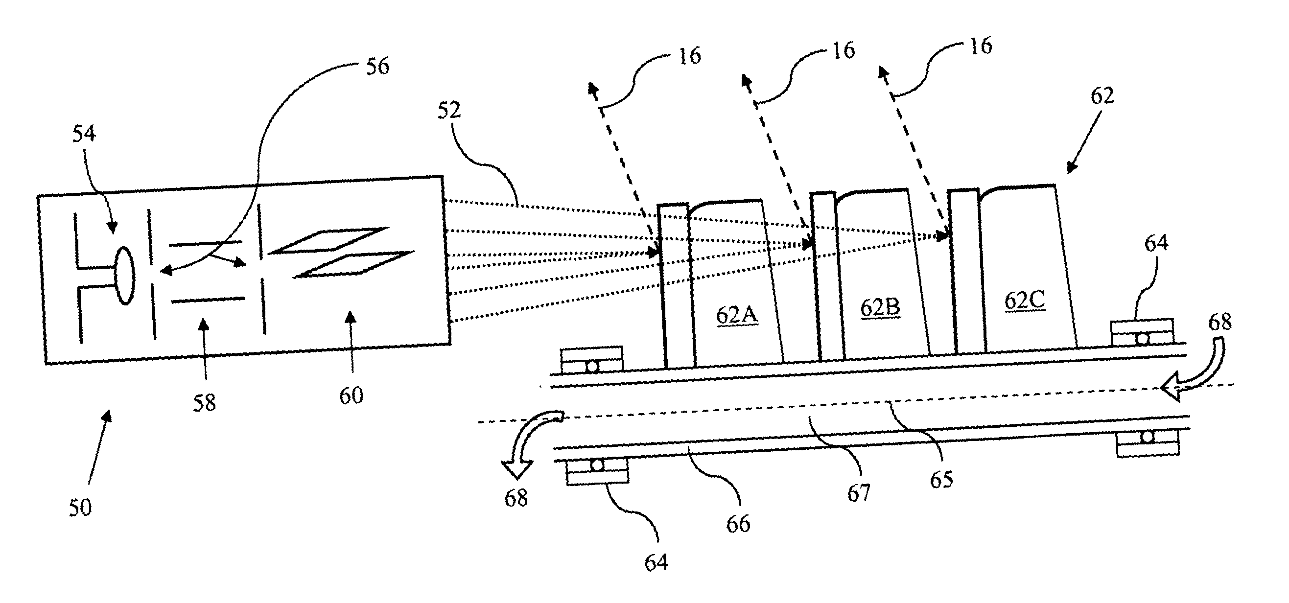
[0006] Accordingly, the above-identified shortcomings of existing CT and VCT imaging systems and methods are overcome by embodiments of the present invention, which relates to a single, near-linear, multi-spot x-ray source comprising multiple targets that have varying focal spots thereon. Embodiments of this invention allow thicker multi-slice images (up to about 80-160 mm thick or thicker) to be obtained with each gantry rotation than currently possible with existing CT and VCT imaging systems.

Problems solved by technology

Currently, x-ray sources for CT and VCT are limited to fairly narrow “slices” for each revolution of the gantry because of the well-understood “cone-beam artifact” problem, in which the “edges” of the cone-like x-ray beam that emerges from a point source cannot produce enough attenuation data, thereby resulting in portions of the imaged object not being imaged at all.

Method used


Image

  • Extended multi-spot computed tomography x-ray source
  • Extended multi-spot computed tomography x-ray source
  • Extended multi-spot computed tomography x-ray source

Examples


Embodiment Construction

[0020] For the purposes of promoting an understanding of the invention, reference will now be made to some preferred embodiments of the present invention as illustrated in FIGS. 1-5 and specific language used to describe the same. The terminology used herein is for the purpose of description, not limitation. Specific structural and functional details disclosed herein are not to be interpreted as limiting, but merely as a basis for the claims as a representative basis for teaching one skilled in the art to variously employ the present invention. Any modifications or variations in the depicted support structures and methods, and such further applications of the principles of the invention as illustrated herein, as would normally occur to one skilled in the art, are considered to be within the spirit and scope of this invention.

[0021] This invention relates to systems and methods for minimizing or eliminating the cone-beam artifact problem in CT images, particularly VCT images, to all...


PUM

PropertyMeasurementUnit
total thicknessaaaaaaaaaa

Abstract

Systems and methods for obtaining multi-slice images having a total thickness of up to about 160 mm or more in a single gantry rotation in computed tomography or volume computed tomography are described. One embodiment comprises an extended, multi-spot x-ray source for computed tomography or volume computed tomography imaging, comprising: an electron gun capable of producing a plurality of electron beams, each electron beam focused at a predetermined distance and aimed in a predetermined direction; and a plurality of targets positioned to receive the electron beams and generate x-rays in response thereto, each target comprising a predetermined focal spot thereon, wherein each electron beam is synchronized to strike, at an appropriate time, a predetermined target comprising a predetermined focal spot thereon.

Description

FIELD OF THE INVENTION [0001] The present invention relates generally to computed tomography (CT) imaging and volume computed tomography (VCT) imaging. More specifically, the present invention relates to multi-spot x-ray sources for CT imaging. Even more specifically, the present invention relates to a stand-alone, self-contained electron gun, having electron beams focusable at different distances, which impinge on multiple targets to generate near-linear multi-spot x-rays for CT and VCT imaging. BACKGROUND OF THE INVENTION [0002] Computed tomography (CT), sometimes called computed axial tomography (CAT) or CAT scan, and volume computed tomography (VCT), use special x-ray equipment to obtain image data from different angles around a person's body, and then use computer processing of the data to create a two-dimensional cross-sectional image (i.e., slice) or three-dimensional image of the body tissues and organs that were scanned. CT and VCT imaging are particularly useful because th...

Claims


Application Information

Patent Timeline
Patent Type & Authority Applications(United States)
IPC IPC(8): A61B6/03G01N23/06G21G4/00H01J35/00H01J35/08H01J35/14H05G1/02H05G1/26H05G1/60H05H1/00
CPCH01J35/08H01J2235/086H01J35/14H01J35/153H01J35/10H01J35/147
Inventor PRICE, JOHN SCOTTBLOCK, WAYNE FREDERICKVERMILYEA, MARK ERNEST
Owner GE MEDICAL SYST GLOBAL TECH CO LLC
Why Eureka
Eureka Blog
Learn More