
Image guided radiosurgery method and apparatus using registration of 2D radiographic images with digitally reconstructed radiographs of 3D scan data

a radiosurgery and image registration technology, applied in the field of radiosurgery, can solve the problems of difficult to meet both requirements simultaneously, difficult to establish, and the skeletal structure of drr images cannot be reconstructed very well, and achieve the effect of accurate and rapid method and system

Active Publication Date: 2005-03-03
ACCURAY
View PDF23 Cites 188 Cited by
  • Summary
  • Abstract
  • Description
  • Claims
  • Application Information

AI Technical Summary

Benefits of technology

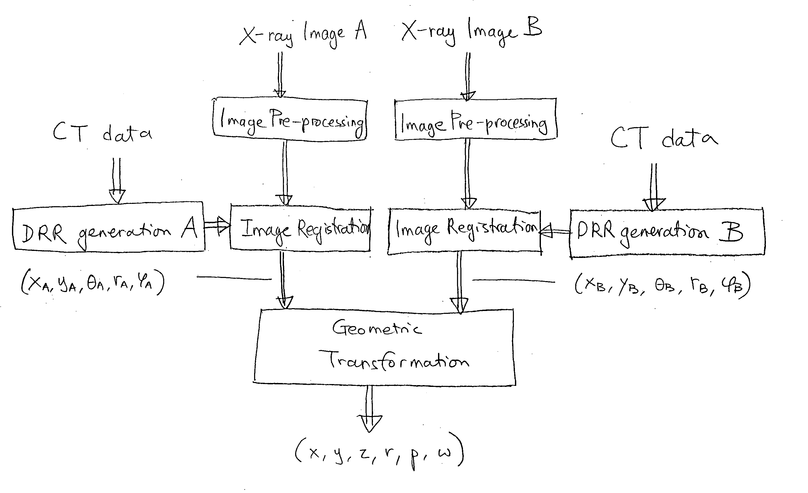
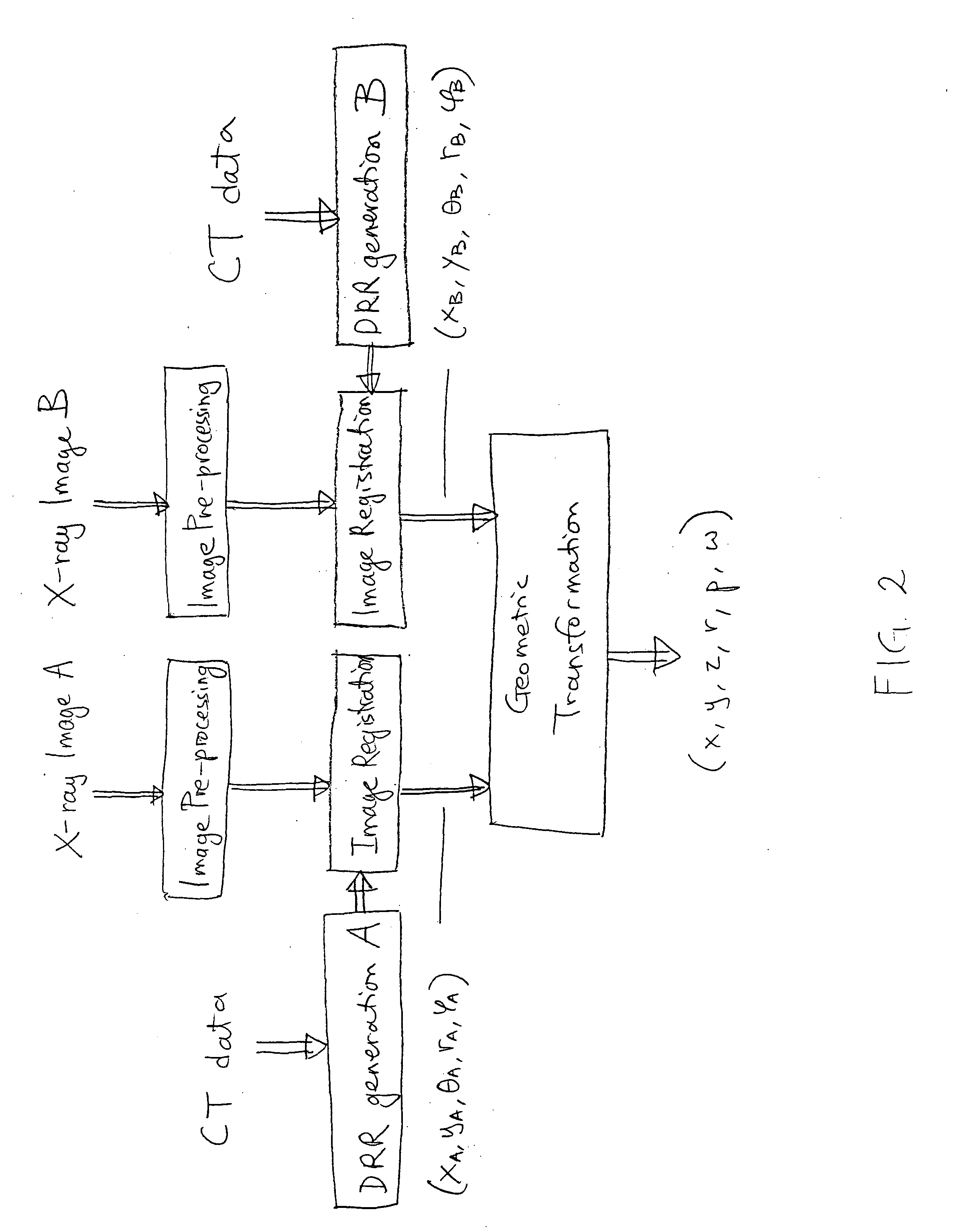
[0009] The present invention is directed to an accurate and rapid method and system for tracking target position for image guided radiosurgery. A hierarchical and iterative 2D / 3D registration algorithm is used that allows for accurate and rapid correction of target position, and alignment of radiosurgical beams, throughout the treatment procedure.

Problems solved by technology

Unfortunately, it is difficult to meet both requirements simultaneously, because of a number of reasons.
It is difficult to establish this relationship, however, so the linear attenuation model is often used.
The skeletal structures in DRR images cannot be reconstructed very well when the linear model is used.
However, the full resolution of the x-ray images is rarely used, because of the extremely slow computation time that results from such use.
Because the full information content of the image is not used, however, accuracy is sacrificed.
Because a long time computation is required, however, image intensity based methods are not practical for radiosurgery, or for clinical practice in general.
However, the accuracy and stability of the estimates are not sufficient to meet the sub-mm precision that is required in radiosurgery applications.

Method used


Image

  • Image guided radiosurgery method and apparatus using registration of 2D radiographic images with digitally reconstructed radiographs of 3D scan data
  • Image guided radiosurgery method and apparatus using registration of 2D radiographic images with digitally reconstructed radiographs of 3D scan data
  • Image guided radiosurgery method and apparatus using registration of 2D radiographic images with digitally reconstructed radiographs of 3D scan data

Examples


Embodiment Construction

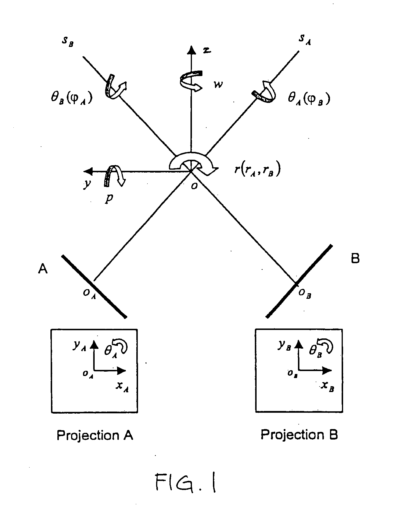
[0017] The present invention features an accurate and rapid method and system for tracking the position of a treatment target in image-guided radiosurgery. The position of the target is tracked in six degrees of freedom. The present invention is useful for patient position correction and radiation beam alignment during radiosurgery / radiotherapy of a treatment target, for example a tumor within the brain or skull. The method and system of the present invention allows for a fully automatic tracking process, with no need for user interaction.

[0018] In overview, 2D reconstructed radiographs (typically, DRRs) are generated offline from a pre-treatment 3D scan. The 3D scan is typically a CT scan, however, any other 3D diagnostic scan (such as MRI or PET) may also be used. The DRRs are used as references for determining the position of the target. X-ray images are then acquired in near real time during treatment, in order to determine the change in target position. A hierarchical and iter...


PUM


Abstract

An image-guided radiosurgery method and system are presented that use 2D / 3D image registration to keep the radiosurgical beams properly focused onto a treatment target. A pre-treatment 3D scan of the target is generated at or near treatment planning time. A set of 2D DRRs are generated, based on the pre-treatment 3D scan. At least one 2D x-ray image of the target is generated in near real time during treatment. The DRRs are registered with the x-ray images, by computing a set of 3D transformation parameters that represent the change in target position between the 3D scan and the x-ray images. The relative position of the radiosurgical beams and the target is continuously adjusted in near real time in accordance with the 3D transformation parameters. A hierarchical and iterative 2D / 3D registration algorithm is used, in which the transformation parameters that are in-plane with respect to the image plane of the x-ray images are computed separately from the out-of-plane transformation parameters.

Description

FIELD OF THE INVENTION [0001] The present invention relates to radiosurgery. In particular, the present invention relates to an image guided radiosurgery method and apparatus using 2D / 3D image registration. BACKGROUND [0002] Radiosurgery is used to treat tumors and other lesions by delivering a prescribed high dose of high-energy radiation to the target area while minimizing radiation exposure to the surrounding tissue. In radiosurgery, precisely focused beams of radiation (e.g. very intense x-ray beams) are delivered to a target region in order to destroy tumors or to perform other types of treatment. The goal is to apply a lethal amount of radiation to one or more tumors, without damaging the surrounding healthy tissue. Radiosurgery therefore calls for an ability to accurately target a tumor, so as to deliver high doses of radiation in such a way as to cause only the tumor to receive the desired dose, while avoiding critical structures such as the spinal cord. [0003] Conventional ...

Claims


Application Information

Patent Timeline
IPC IPC(8): A61N5/10G01N23/04G06T7/00G06T7/20
CPCA61N5/1049A61N5/1067A61N5/107A61N2005/1061A61N2005/1062G06T2207/30004G06T7/0026G06T7/0038G06T7/2053G06T2207/10124G01N23/04G06T7/32G06T7/38G06T7/254
Inventor KUDUVALLI, GOPINATHFU, DONGSHANQURESHI, SHEHRZAD
Owner ACCURAY
Learn More