
X-ray image diagnostic device, and x-ray image data correcting method

Inactive Publication Date: 2004-12-02
HITACHI LTD
View PDF3 Cites 23 Cited by
  • Summary
  • Abstract
  • Description
  • Claims
  • Application Information

AI Technical Summary

Benefits of technology

[0007] Therefore, an object of the present invention is to provide an X-ray image diagnostic device capable of obtaining sensitivity information for various X-ray intensities from a single output image and performing accurate sensitivity correction.

Problems solved by technology

This causes a problem in that sensitivity correction is not accurate.
This causes another problem in that the work of the operator for collecting sensitivity information increases and that the capacity of the storage medium for storing the collected sensitivity information must be increased.

Method used


Image

  • X-ray image diagnostic device, and x-ray image data correcting method
  • X-ray image diagnostic device, and x-ray image data correcting method
  • X-ray image diagnostic device, and x-ray image data correcting method

Examples


Embodiment Construction

[0018] Embodiments of the X-ray image diagnostic device of the present invention will be explained with the reference to the attached drawings.

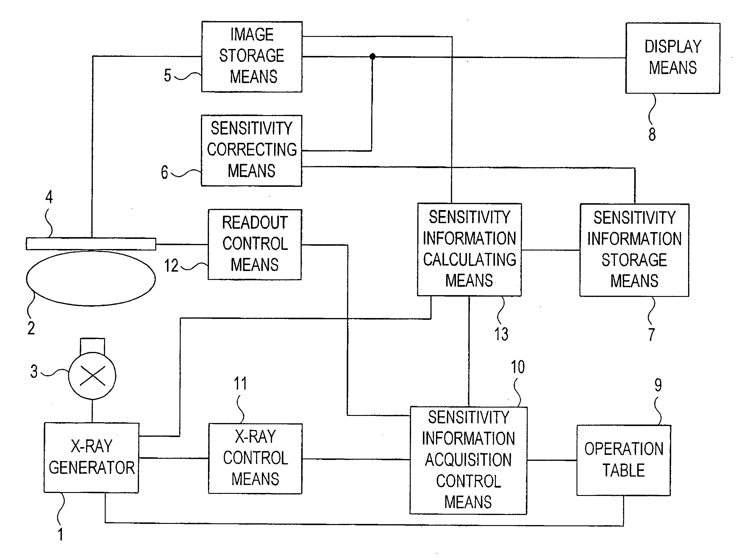
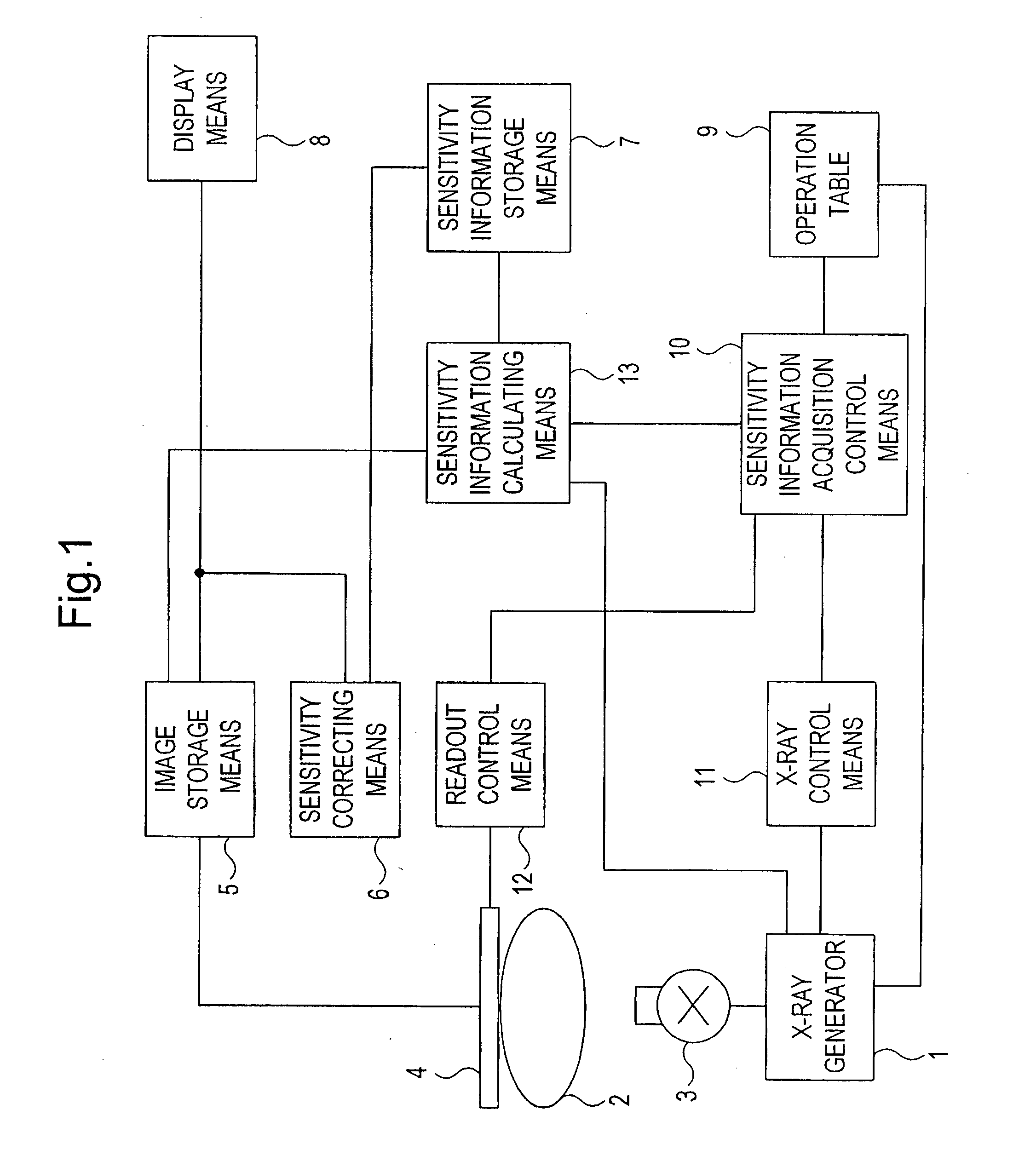
[0019] FIG. 1 is a functional diagram showing the X-ray image diagnostic device of the present invention.

[0020] As shown in FIG. 1, the X-ray image diagnostic device of the present invention has an X-ray source 3 which irradiates X-rays onto an object 2 to be imaged under the control of an X-ray generator 1, and an X-ray plane detector which is placed face to face with the X-ray source 3 and outputs transmitted X-rays through the object 2 as X-ray image data. The X-ray image diagnostic device further comprises means for displaying X-ray image data output from the X-ray plane detector 4 as images (5-8), control means for controlling X-ray irradiation and image-signal readout from the X-ray plane detector 4 (10,11,12), and an operation table 9 for inputting directions and conditions necessary for operation of the device etc.

[0021] The X-ray gen...


PUM


Abstract

An X-ray image diagnostic device of the invention has sensitivity-information-acquisition controlling means commanding synchronization between X-ray irradiation from the X-ray source and readout of X-ray plane detector for correcting sensitivity. When sensitivity information of the X-ray plane detector is calculated using the intensities of X-rays irradiated onto the X-ray plane detector and X-rays detected by the X-ray plane detector, readout of the X-ray plane detector is performed synchronously with X-ray irradiation, and sensitivity information corresponding to variation of the amount of X-ray radiation onto the X-ray plane detector during the reading time is determined using data of every readout channel. Therefore, sensitivity information enabling an accurate sensitivity correction can be obtained from a single output image. This sensitivity information is stored in sensitivity-information-storing means and used for correcting sensitivity of X-ray image data acquired in actual measurements.

Description

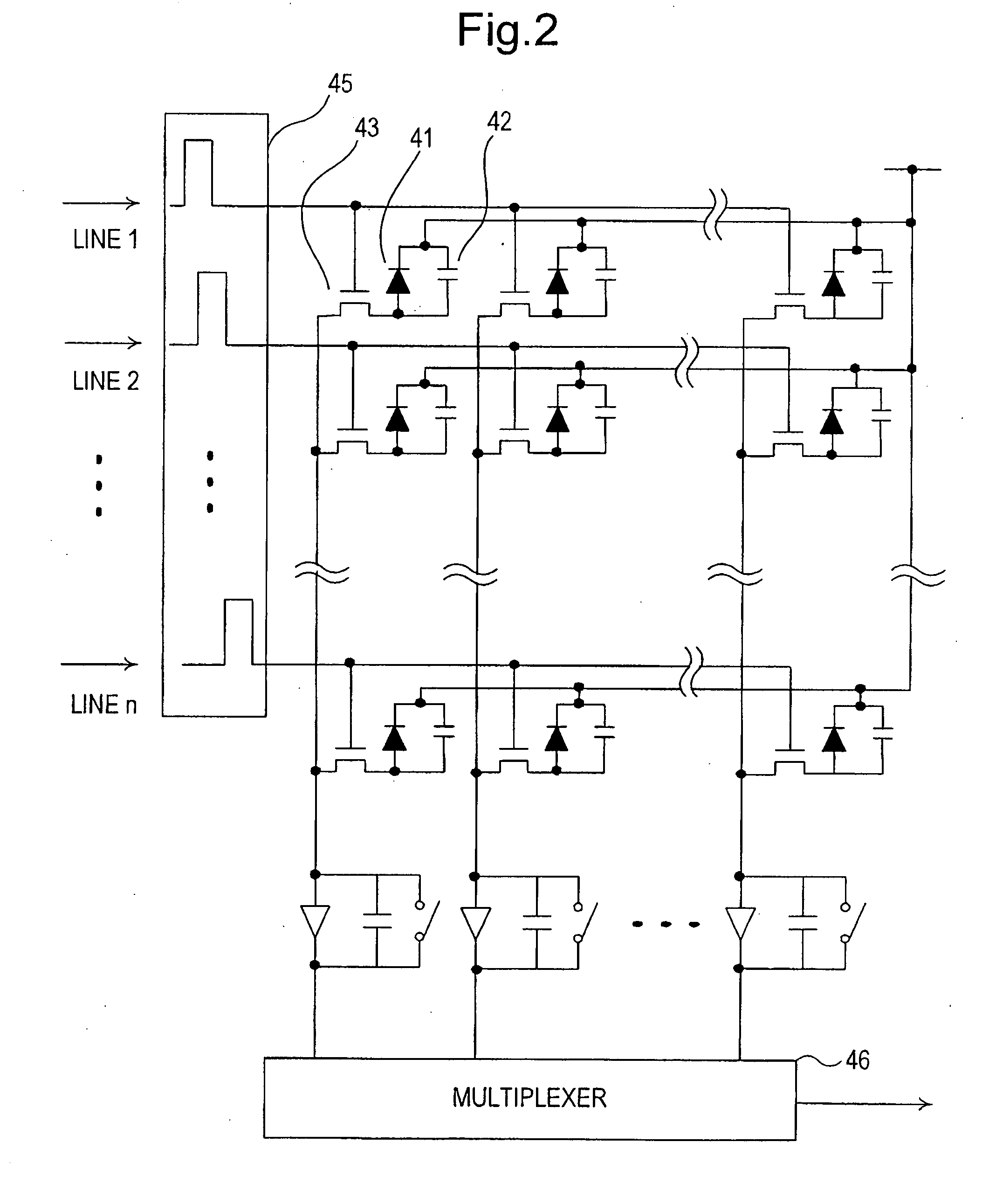
[0001] The present invention relates to an X-ray image diagnostic device using an X-ray plane detector. Particularly, it relates to an X-ray image diagnostic device having a function of correcting sensitivity to X-ray energy of an X-ray plane detector exposed to X-rays.RELATED ART[0002] Conventional X-ray image diagnostic devices are constructed such that an X-ray image output from an X-ray plane detector resulting from irradiation of X-rays onto an object is displayed on a TV monitor or the like.[0003] An ordinary X-ray plane detector consists of scintillators that convert X-rays transmitted through an object to light and photodiodes that convert the light output from the scintillators to electric charge. Detecting devices, each of which consists of a scintillator and a photodiode, are arranged in a matrix corresponding to the individual pixels. Electric charge converted from light by each photodiode is read out via a switching device such as a thin film transistor (TFT) to produce...

Claims


Application Information

Patent Timeline
IPC IPC(8): A61B6/00
CPCA61B6/542A61B6/583A61B6/585
Inventor SUZUKI, KATSUMI
Owner HITACHI LTD
Learn More