
Method for testing a cell sample

a cell sample and cell technology, applied in the field of cell sample testing, can solve the problems of misdiagnosis and patient's death, method most often fails, and error is rarely recognised

Inactive Publication Date: 2002-03-28
SHINE THOMAS ADAM +1
View PDF0 Cites 10 Cited by
  • Summary
  • Abstract
  • Description
  • Claims
  • Application Information

AI Technical Summary

Benefits of technology

[0027] Although it is possible for the method of the invention to comprise merely of the treatment of two aliquots of the sample cell suspension, more usually the method includes the steps of subjecting another aliquot of sample cell suspension to a second alteration in at least one parameter of the cell environment passing said altered aliquot through the sensor, recording the change in said property of the cell suspension under the altered environment as each of a number of cells of the aliquot passes through the sensor, recording all the concomitant properties of the environment together with the said change on a cell-by-cell basis, and comparing the data from previous step (c) and the preceding step as a function of the extent of said second alteration of environmental parameter. Usually there are many further aliquots treated in a similar way. The greater the number of aliquots tested, the greater the potential accuracy, precision and resolution of the results which are obtained. It is also possible to subject a only single aliquot of sample suspension to a series of such alterations in at least one parameter of the cell environment.
[0032] In current practice, cell shape, particularly red blood cell shape, is not estimated by any automated method. The present invention enables the user to determine cell shape and derive other data, such as cell volume, surface area, surface area to volume ratio, sphericity index, cell thickness, and surface area per milliliter. Aside from research and experimental laboratories, none of these measurements are currently available in any clinical laboratory and hitherto, none could be completed within 60 seconds. In particular, the preferred method where the sample cell suspension is subjected to a concentration gradient, enables the automatic detection or a user to detect accurately when the cells adopt a substantially spherical shape immediately before lysis.
[0034] In the preferred method of the present invention, this fixed shape correction factor is replaced with a sample specific shape correction factor f(K.sub.shape), generated from a shape correction function. The shape correction function is continuous for all cell shapes and ranges in value from 1.0 for spherical cells to infinity for a perfectly flat cell. The shape correction function increases the accuracy with which cell parameters which depend on anatomical measurement, such as cell volume, can be determined.
[0050] The present invention provides a method for testing blood samples which enables data to be obtained on a cell-by-cell basis. By using the data on a cell-by-cell basis, it enables new parameters to be measured and to obtain information on the distribution of cells of different sizes among a population and reveal sub-populations of cells based on their anatomical and physiological properties.
[0089] This list gives an indication of the clinical value of the present invention which is greatly enhanced once the changes can be quantified and the subtler changes are associated with disease. The ability to measure the degree of thickness or thinness of cells allows the possibility to study the progress of the disease and assess the value of treatment, especially at an early stage.

Problems solved by technology

In the majority of patients, these results do not cause significant problems but occasionally the erroneous measurements result in misdiagnosis and patient's death.
However, this error is rarely recognised because existing automated methods cannot detect it.
It is these abnormal samples that indicate pathology where accuracy is most needed that the method most often fails.

Method used


Image

  • Method for testing a cell sample
  • Method for testing a cell sample
  • Method for testing a cell sample

Examples


Embodiment Construction

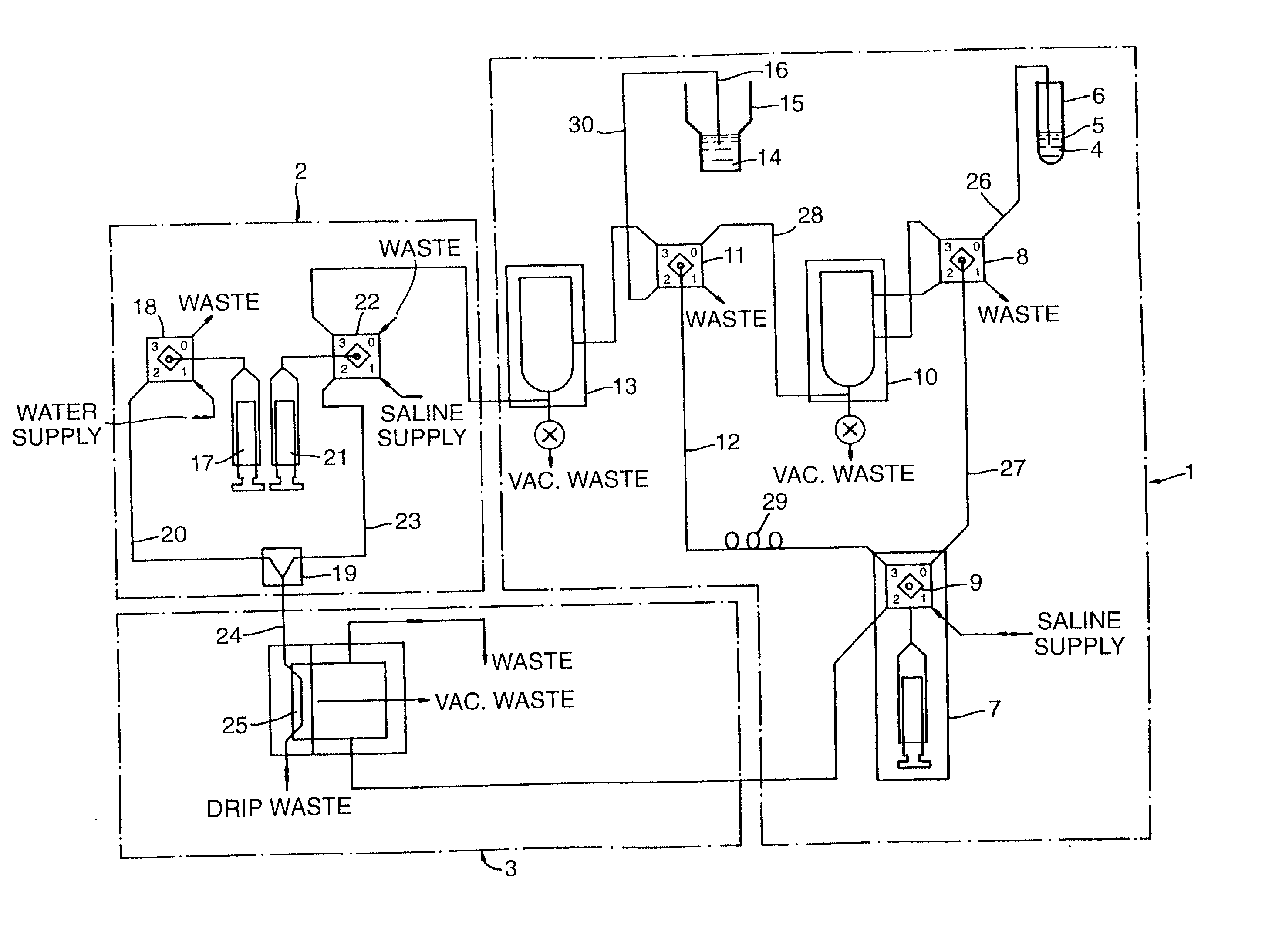
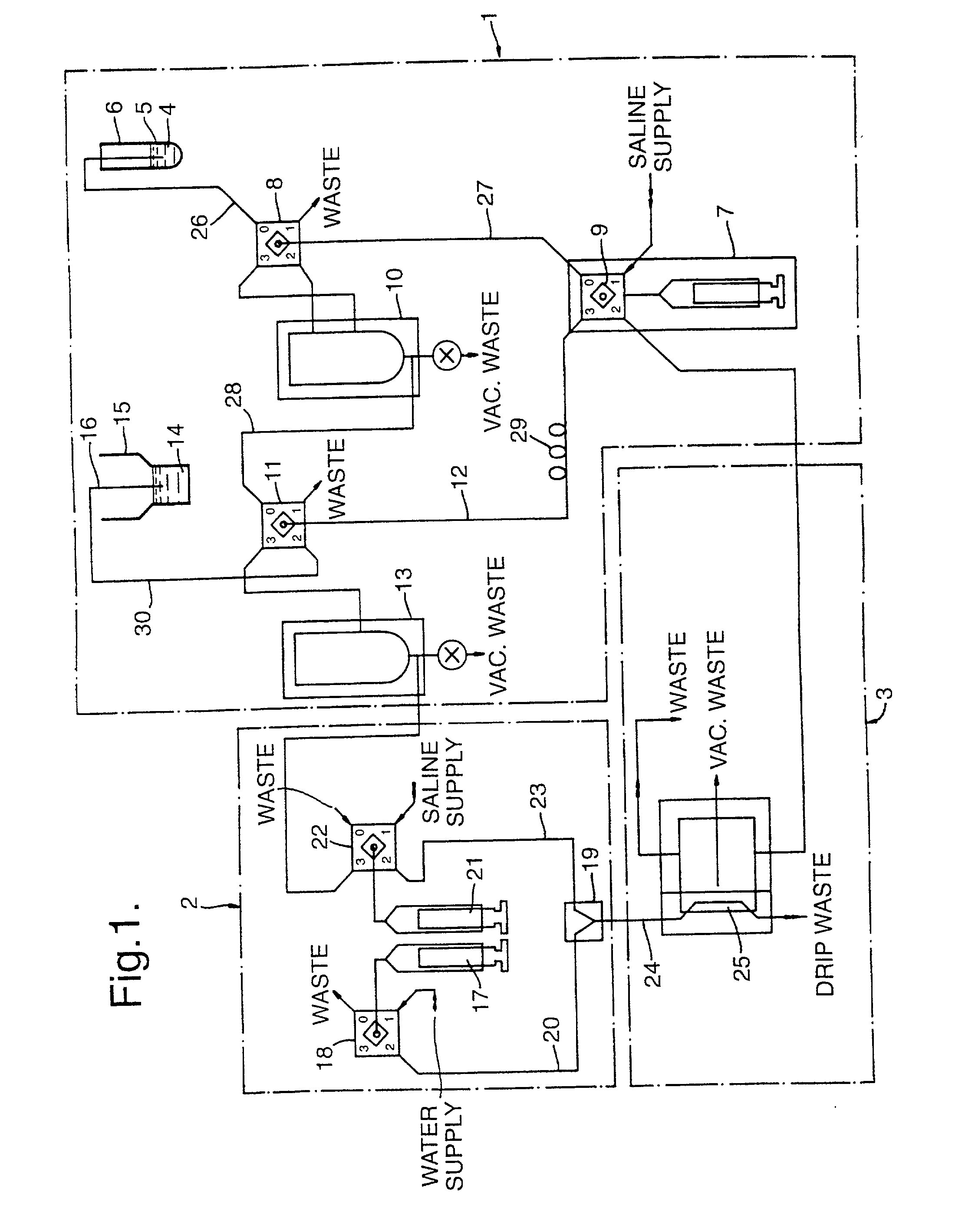
[0092] FIG. 1 shows schematically an instrument used to sample and test blood cells;

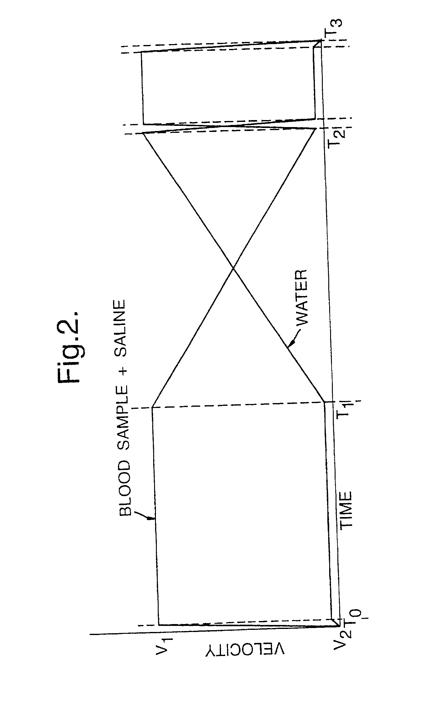
[0093] FIG. 2 shows velocity profiles for the discharge of fluids from fluid delivery syringes of a gradient generator section of the instrument of FIG. 1;

[0094] FIG. 3 shows a block diagram illustrating the data processing steps used in the instrument of FIG. 1;

[0095] FIG. 4 shows an example of a three-dimensional plot of osmolality against measured voltage for cells of a blood sample analyzed in accordance with the present invention;

[0096] FIG. 5 shows another example of a three-dimensional plot of osmolality against measured voltage which illustrates the frequency distribution of blood cells at intervals;

[0097] FIG. 6 shows a series of three-dimensional plots for a sample tested at hourly intervals;

[0098] FIGS. 7 and 8 show results for spherical latex particles as part of an instrument calibration routine;

[0099] FIG. 9 shows superimposed plots of osmolality (x-axis) against measured voltage and tr...


PUM


Abstract

In this invention, a cell parameter, for example cell volume, is determined by subjecting one or more aliquots of a sample cell suspension to one or more alterations of at least one parameter of the cell environment to identify a point at which the cells achieve a particular shape to obtain a sample specific shape compensation factor. Preferably, the environmental parameter change is a reduction in osmolality.

Description

[0001] This invention relates to a method for testing a cell sample or a fluid sample.[0002] Many types of cells as well as artificial cells may be tested according to the method of the present invention, although the test is especially suitable for testing red and white blood cells.[0003] It is well known that certain diseases give rise to changes in the condition of blood and especially of the red blood cells. Blood characteristics such as red blood cell count, mean cell volume, haemoglobin content and haematocrits are commonly used in the diagnosis of disease.[0004] The measurement of cell volume is one of the most informative investigations in clinical medicine. Cell size is an important indicator of pathology and in conjunction with haemoglobin, forms the basic classification of anemias. It is the basis of the differentiation of white blood cells and leukaemias, the assessment of prognosis in scores of diseases and is an invaluable screening device for the thalassaemias, the mo...

Claims


Application Information

Patent Timeline
IPC IPC(8): G01N15/00G01N15/12G01N33/49
CPCG01N15/12G01N2015/0073G01N2015/105G01N2015/1062G01N2015/1087G01N2015/1093G01N2015/012G01N2015/1022G01N2015/103G01N2015/1024G01N2015/1029
Inventor SHINE, THOMAS ADAMSHINE, IAN BASIL
Owner SHINE THOMAS ADAM
Learn More