
Noninvasive detection of colorectal cancer and other gastrointestinal pathology

Inactive Publication Date: 2001-09-27
NAIR PADMANABHAN P
View PDF0 Cites 5 Cited by
  • Summary
  • Abstract
  • Description
  • Claims
  • Application Information

AI Technical Summary

Benefits of technology

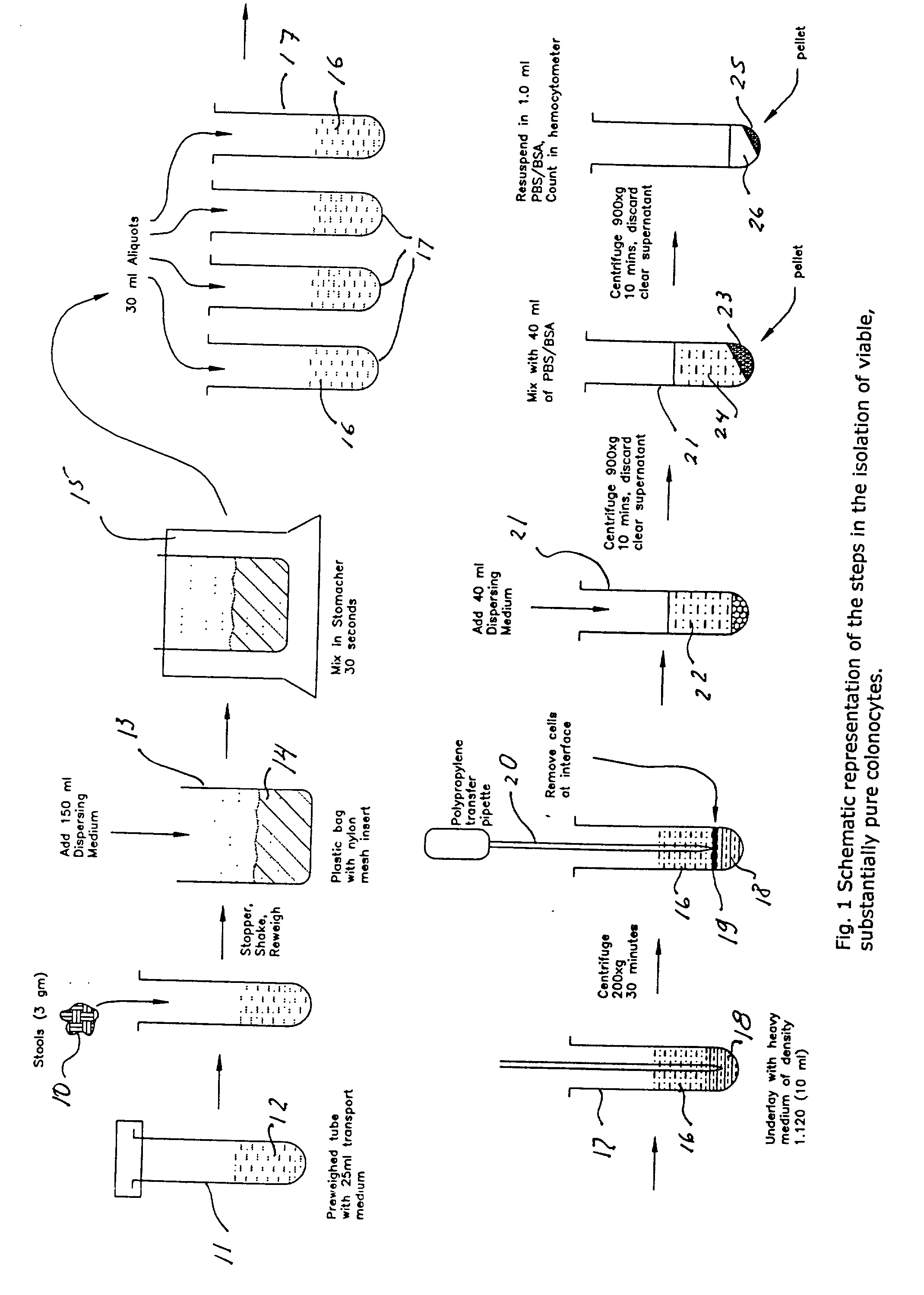
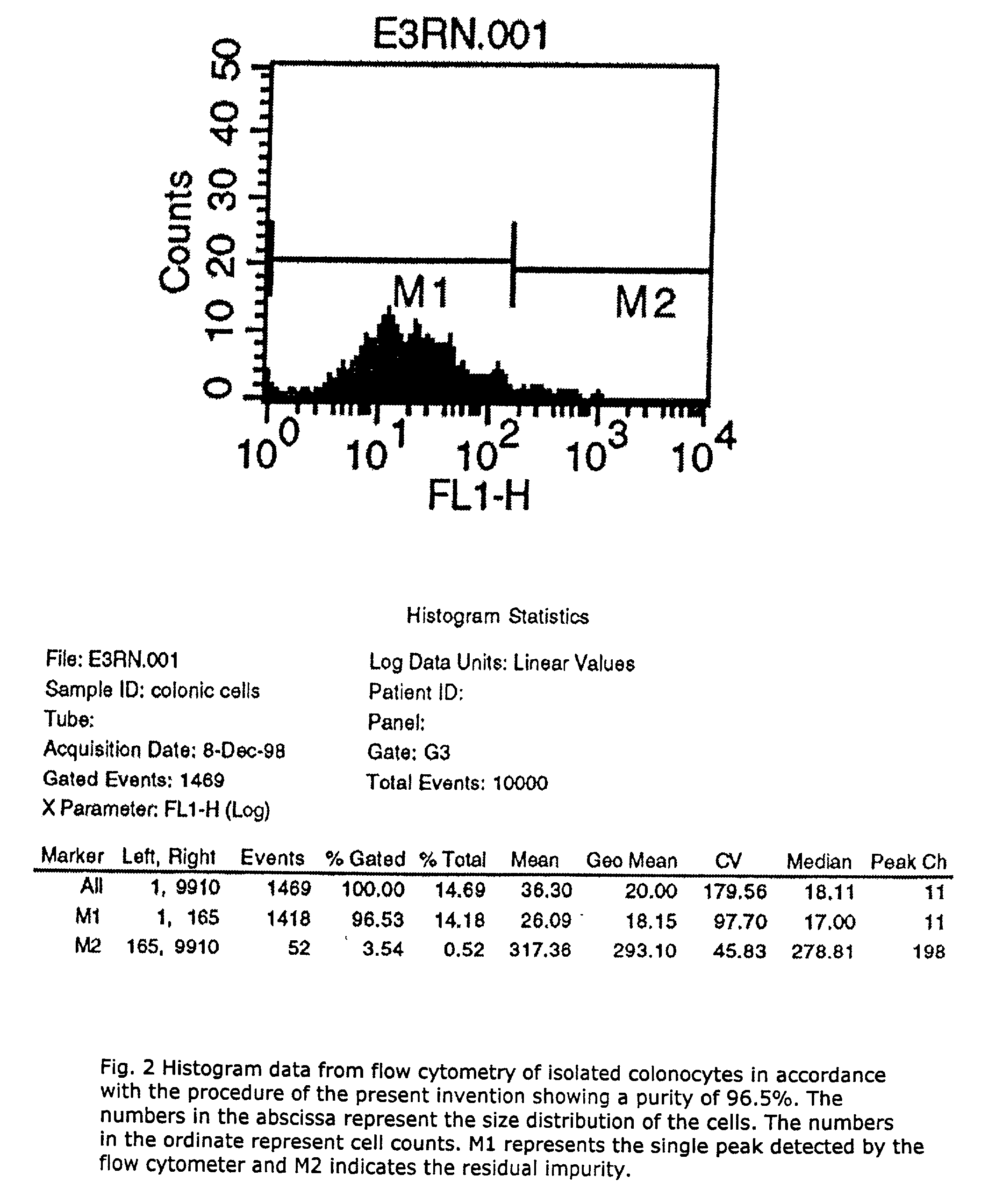
[0036] It is important to note that an inventive aspect of the present invention is the formulation of a transport medium and a dispersion or suspension medium which together allow the exfoliated colonocytes found in the fecal sample to remain viable at the normal ambient temperature during and after the isolation procedure. In other words, the invention enables isolation of intact, living exfoliated colonocytes from a small sample (e.g. 3 gm) of fecal material without chilling or freezing, at normal ambient temperature ranging from about 22.degree. C. to about 24.degree. C. during the entire isolation process. About 8 to 10 million living colonic cells can be obtained from one gram of the stool sample in accordance with the techniques of the present invention.

Problems solved by technology

However, frequent false positive and false negative results of the FOBT considerably limit its specificity and sensitivity.
Other procedures are expensive and invasive.
Thus, these procedures do not retain cellular integrity and are not applicable for isolating intact, viable cells free of other impurities.
Thus, the procedures of Iyengar et al and Albaugh et al may provide viable colonocytes, but they are inadequate for the purpose of obtaining substantially pure colonocytes.
In summary, heretofore it has not been possible to obtain a viable, biologically subtantially pure sample of a particular cell type isolated at normal ambient atmospheric conditions from a small fecal mass.

Method used


Image

  • Noninvasive detection of colorectal cancer and other gastrointestinal pathology
  • Noninvasive detection of colorectal cancer and other gastrointestinal pathology
  • Noninvasive detection of colorectal cancer and other gastrointestinal pathology

Examples


example-2

Assessment of Status of Mucosal Immunity

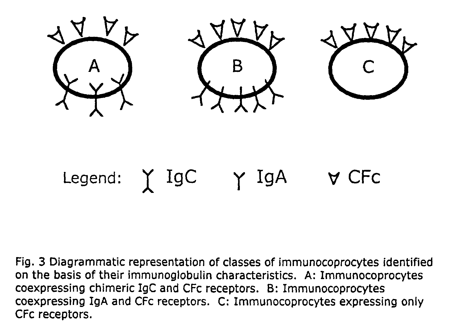
[0052] Cells are obtained as described above from subjects who are suspected of having an immunocompromised gut. Aliquots of cells (about 110K) are suspended in PBS buffer and incubated at 37.degree. C. for about 45 minutes with one of the following combinations of fluorescently labelled antibodies: anti-IgG FITC (green), anti-IgA PE (red), and anti-IgC FITC+anti-IgA PE. A parallel tube containing cells with an isotype control antibody is also maintained to account for non-specific binding antibody. Direct immunofluorescence assays are conducted to measure the binding of antibodies to different sets of colonic cells. A significant decrease in number of immunocoprocytes (expressing IgC) or IgA bearing cells is of diagnostic significance for immune deficiency. Table 2 lists values obtained for normal subjects. Any deviation from the normal values would be indicative of immune dysfunction. It should be noted that direct and indirect immunofluores...

example-3

Expression of Colon Cancer-Associated Biomarkers

[0053] Cells are obtained as described above from patients suspected of having colon cancer or precursors of colon cancer (polyps). In one embodiment of this technique, cells are subjected to indirect immunofluorescence assay for the expression of CD44 or its molecular variants, e.g., CD44V3, CD44V6 and CD44V10; the presence of CD44 or its molecular variants being diagnostic of colon cancer.

example-4

[0054] As a source of somatic cells obtainable non-invasively, the colonocytes of the present invention are representative of the phenotype as well as genotype. Thus, they are useful in DNA typing and examination of biological macromolecules (such as DNA, RNA, protein and the like) for determining responses, for example, to pharmacologic and environmental agents and assessment of multi-drug resistance. These isolated cells are also useful in various other ways easily suggested to a skilled artisan.

[0055] It should be apparent that given the guidance, illustrations and examples provided herein, various alternate embodiments, modifications or manipulations of the present invention would be suggested to a skilled artisan and these are included within the spirit and purview of this application and scope of the appended claims.

2TABLE 1 Effect of storage on cell yields and viability Storage Time Cell Yield (Hrs) Conditions % Viability 1-6 Ambient 100 85+ 10 Ambient 100 65 24 4.degree. C. ...


PUM

PropertyMeasurementUnit
Massaaaaaaaaaa
Massaaaaaaaaaa
Massaaaaaaaaaa

Abstract

A method for isolating viable, biologically substantially pure exfoliated fecal colonocytes at normal ambient temperature is described. Immunocoprocytes and inflammatory cells indicative of certain gastrointestinal conditions and a noninvasive method for detecting colorectal cancer are set forth. Composition of transport and suspension media for isolation of colonocytes are detailed.

Description

[0001] 1. Field of the Invention[0002] The present invention is related to isolated colonocytes enabling early noninvasive detection of colorectal cancer and other gastrointestinal diseases. More particularly, the present invention is related to isolated, biologically substantially pure and viable immunocoprocytes and nonepithelial cells of lymphoid origin obtained from a small fecal sample. The invention is further related to providing a transport medium and a dispersion or suspension medium for isolating viable colonocytes from a fecal sample at normal ambient temperature and a method for detecting colorectal and other gastrointestinal pathology employing the isolated colonocytes of the present invention. The isolated colonocytes also allow the study and determination of other anomalous conditions, symptoms, disorders or pathological conditions.[0003] 2. Prior Art[0004] A common gastrointestinal malignancy in humans is colorectal cancer. It has been estimated that colorectal cance...

Claims


Application Information

Patent Timeline
IPC IPC(8): C07K16/00C07K16/40C12N5/02C12N5/071C12N5/09G01N33/574G01N33/68
CPCC07K16/00C07K16/40C12N5/0679C12N5/0693G01N33/57419G01N33/6893G01N2333/90241G01N2800/06C07K2317/52
Inventor NAIR, PADMANABHAN P.
Owner NAIR PADMANABHAN P
Learn More