Soft tissue matrix characterization using stretched exponential relaxation modeling

a stretching and relaxation modeling technology, applied in the field of magnetic resonance imaging, can solve problems such as neck and back pain, and achieve the effects of reducing error, reducing neck and back pain, and reducing sensitivity to specific matrix components

Active Publication Date: 2021-09-28
UNIV OF COLORADO THE REGENTS OF +1
View PDF8 Cites 0 Cited by
  • Summary
  • Abstract
  • Description
  • Claims
  • Application Information

AI Technical Summary

Benefits of technology

[0006]Intervertebral disc degeneration (IVDD), resulting in the depletion of hydrophilic glycosaminoglycans (GAGs) located in the nucleus pulposus (NP), can lead to debilitating neck and back pain. Magnetic Resonance Imaging (MRI) is a promising means of IVD assessment due to the correlation between GAG content and MRI relaxation values. Furthermore, anomalous (i.e. non-monoexponential) relaxation models have yielded higher sensitivity to specific matrix components compared to conventional monoexponential models. Here, T1ρ and T2 relaxation data were obtained from healthy cervical IVDs, and relaxation data was modeled using both conventional and stretched exponential (SE) decays. Normalized histograms of the resultant quantitative MRI (qMRI) maps were fit with stable distributions. SE models fit relaxation behavior with lower error compared to monoexponential models, indicating anomalous relaxation behavior in healthy IVDs. SE model parameters T1ρ and αT1ρ increased with IVD segment, while conventional monoexponential measures did not vary. The improved model fit and correlation between both SE T1ρ and αT1ρ with IVD level indicates that these parameters are more sensitive biomarkers for detection of GAG content variation.

Problems solved by technology

Intervertebral disc degeneration (IVDD), resulting in the depletion of hydrophilic glycosaminoglycans (GAGs) located in the nucleus pulposus (NP), can lead to debilitating neck and back pain.

Method used


Image

  • Soft tissue matrix characterization using stretched exponential relaxation modeling
  • Soft tissue matrix characterization using stretched exponential relaxation modeling
  • Soft tissue matrix characterization using stretched exponential relaxation modeling

Examples


example 1

Materials and Methods

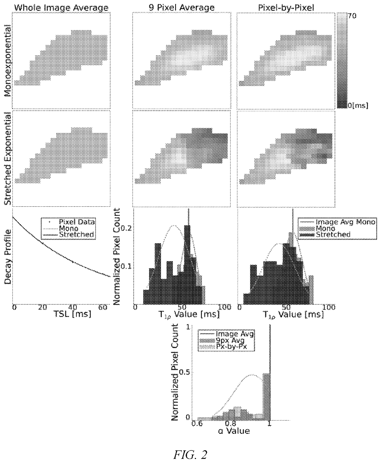
Raw Data Acquisition and Processing

[0029]Cervical IVDs (C2C3-C6C7) were imaged in 15 healthy subjects (7 / 8 males / females; average age=24.7) using a 3T GE MRI scanner. A magnetization-prepared angle-modulated partitioned k-space spoiled gradient echo snapshots (MAPSS) T1ρ sequence was acquired with the following parameters: spin lock power: 500 Hz spin lock times: [1, 5, 20, 40, 60] ms [Li X, Wyatt C, Rivoire J, et al. Simultaneous acquisition of T1ρ and T2 quantification in knee cartilage: Repeatability and diurnal variation. J. Magn. Reson. Imaging 2014; 39:1287-1293 doi: 10.1002 / jmri.24253]. A T2 sequence was also acquired (echo times: [6.78, 13.97, 21.15, 42.72] ms). Shared imaging sequence parameters were: FOV: 14 cm, matrix: 256×128, slice thickness: 4 mm, views per segment: 64, TR 1.2 s, number of slices: 26, ARC acceleration factor: 2, number of signal averages: 4.

[0030]The first spin lock time (TSL) for the T1ρ relaxation data was excluded from relaxatio...

example 2

Results

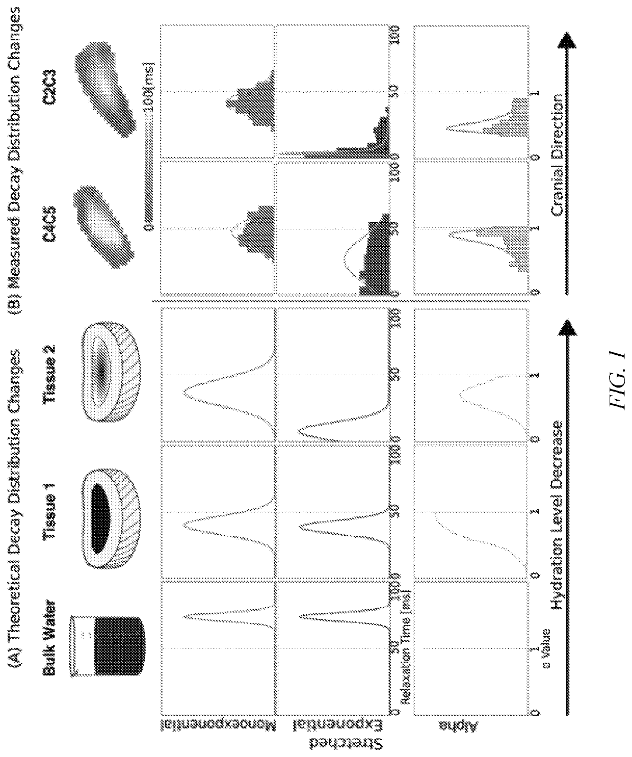
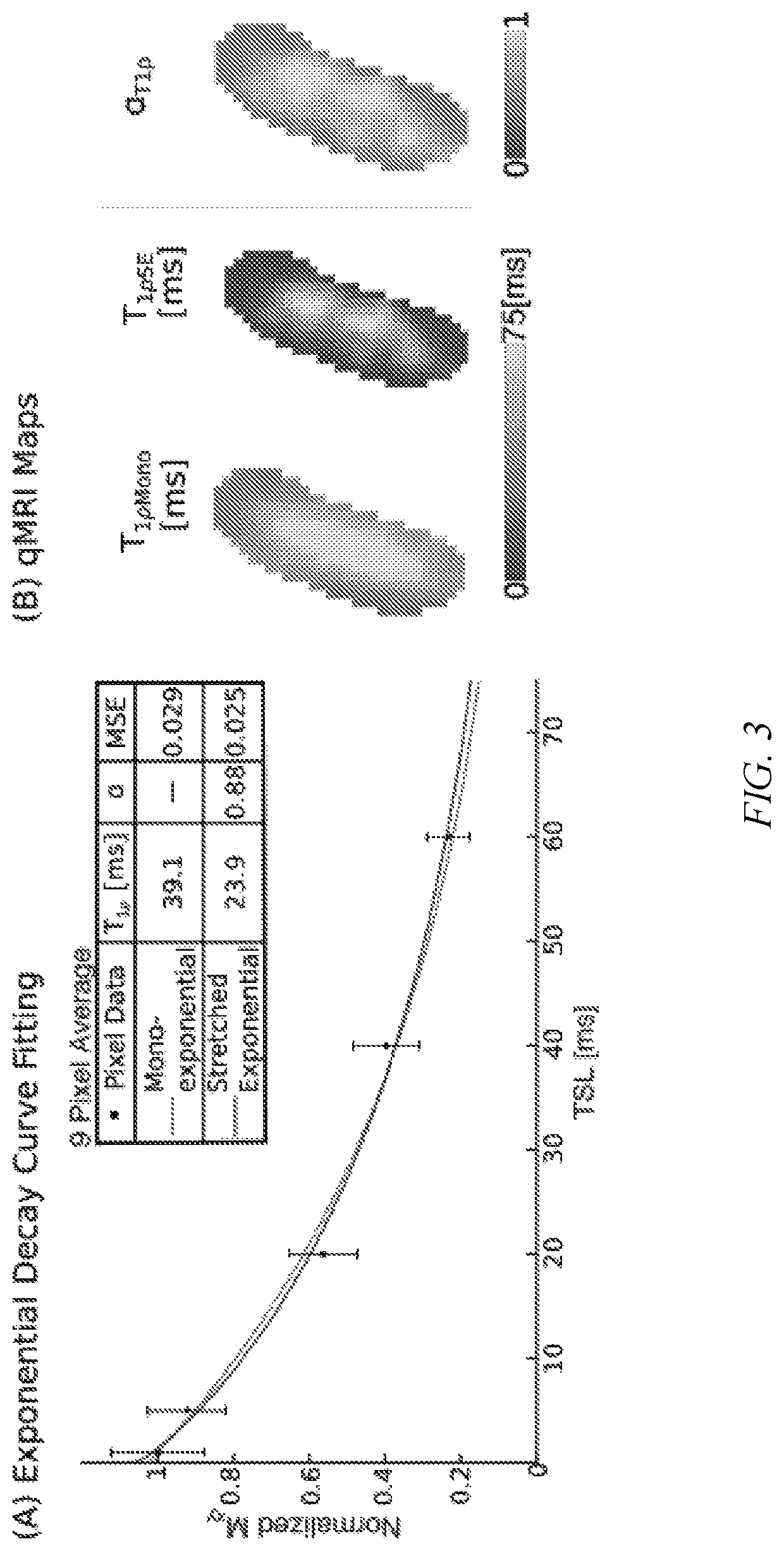
[0034]Single subject analysis shows level-wise increase in SE T2 and T1ρ model parameters moving caudally with IVD segment based on the δ values from the disc-wise distributions, but monoexponential models found no qualitative differences (FIG. 4). MSEs derived from model fits were lower in the stretched exponential model than the conventional model for both T1ρ and T2 (p1ρSE and T2SE) δ values qualitatively increase caudally, a stronger relationship exists in the δ values of the α terms (αT1ρ, αT2). These qualitative observations follow similar trends in both relaxation measures. However, they are more prominent in the T1ρ parameters than T2. The SE (T1ρSE, αT1ρ, T2SE, and αT2) δ values increase at the lower discs (C5C6-C6C7), particularly in the NP compared to the AF (FIGS. 5A and 5B).

[0035]The population level (i) (n=15) T1ρ stable distribution analysis demonstrates a significant relationship between IVD level and SE model parameters (FIG. 6A). Caudally, the shape paramete...


PUM


Abstract

A process for more sensitive characterization of tissue composition for generating a quantitative MRI (qMRI) map and corresponding delta analysis. Intervertebral disc degeneration (IVDD), resulting in the depletion of hydrophilic glycosaminoglycans (GAGs) located in the nucleus pulposus (NP), can lead to debilitating neck and back pain. Magnetic Resonance Imaging (MRI) is a promising means of IVD assessment due to the correlation between GAG content and MRI relaxation values. T1 and T2 relaxation data were obtained from healthy cervical IVDs, and relaxation data was modeled using both conventional and stretched exponential (SE) decays. Normalized histograms of the resultant quantitative MRI (qMRI) maps were fit with stable distributions. SE models fit relaxation behavior with lower error compared to monoexponential models, indicating anomalous relaxation behavior in healthy IVDs. SE model parameters T1 and T1 increased with IVD segment, while conventional monoexponential measures did not vary. The improved model fit and correlation between both SE T1 and T1 with IVD level suggests these parameters are more sensitive biomarkers for detection of GAG content variation.

Description

CROSS-REFERENCE TO RELATED APPLICATIONS[0001]This application claims the benefit of U.S. Provisional Application No. 62 / 695,152, filed Jul. 8, 2018, and U.S. Provisional Application No. 62 / 695,603, filed Jul. 9, 2018.STATEMENT OF GOVERNMENT INTEREST[0002]This invention was made with government support under grant number AR066665, awarded by the National Institutes of Health. The government has certain rights in the invention.FIELD OF INVENTION[0003]This invention relates to magnetic resonance imaging. More specifically, this invention relates to methods of generating quantitative MRI maps for the assessment of intervertebral disc degeneration.BACKGROUND OF THE INVENTION[0004]Intervertebral disc degeneration (IVDD) is a common condition, affecting over 80 million adults in the United States alone, which can lead to debilitating neck and back pain. The intervertebral disc (IVD) structure consists of the collagen-heavy ring-shaped annulus fibrosus (AF) and the glycosaminoglycan (GAG)-r...

Claims


Application Information

Patent Timeline
Patent Type & Authority Patents(United States)
IPC IPC(8): A61B5/00A61B5/055A61B5/0515
CPCA61B5/055A61B5/0515A61B5/4566A61B5/742A61B2560/02
Inventor NEU, COREY P.WILSON, ROBERT L.REITER, DAVID
Owner UNIV OF COLORADO THE REGENTS OF
Why Eureka
Try Eureka