Medical image display device, method, and program

a display device and image technology, applied in image enhancement, instruments, applications, etc., can solve problems such as difficulty in accurately performing comparative observation, and achieve the effects of accurately calculating the change of the case region, accurate performing comparative observation over time, and accurate calculation of the case region

Active Publication Date: 2021-04-20
FUJIFILM CORP
View PDF22 Cites 1 Cited by
  • Summary
  • Abstract
  • Description
  • Claims
  • Application Information

AI Technical Summary

Benefits of technology

[0010]The invention has been made in view of the above circumstances, and it is an object of the invention to make it possible to accurately perform comparative observation over time using images of a disease, such as an interstitial lung disease.
[0025]According to the first medical image display device and method of the invention, a target region included in each of the plurality of medical images having different imaging times for the same subject is classified into a plurality of types of case regions, and a mapping image relevant to each of the case regions corresponding to each of the plurality of medical images is generated by labeling each of the case regions. The center-of-gravity position of a case region is calculated for case regions at corresponding positions in the plurality of mapping images, at least one of the movement amount or the movement direction of the center-of-gravity position between the plurality of mapping images is calculated as a change of each classified case region between the plurality of mapping images, and information regarding the change is displayed on the display unit. As described above, in the invention, since at least one of the movement amount or the movement direction of the center-of-gravity position of each case region at the corresponding position between the plurality of mapping images is calculated as changes of each case region, it is possible to accurately calculate the change of the case region. Therefore, it is possible to accurately perform comparative observation over time using a plurality of medical images having different imaging times.
[0026]According to the second medical image display device and method of the invention, a target region included in each of the plurality of medical images having different imaging times for the same subject is classified into a plurality of types of case regions, and a mapping image relevant to each of the case regions corresponding to each of the plurality of medical images is generated by labeling each of the case regions. The amount of change in the size of a case region at each corresponding position in the plurality of mapping images is calculated as a change of each classified case region between the plurality of mapping images, and information regarding the change is displayed on the display unit. As described above, in the invention, since the amount of change in the size of the case region at each corresponding position is calculated as a change of each case region, it is possible to accurately calculate the change of the case region. Therefore, it is possible to accurately perform comparative observation over time using a plurality of medical images having different imaging times.

Problems solved by technology

Accordingly, it is difficult to accurately perform the comparative observation over time by accurately displaying a region change for each symptom.

Method used


Image

  • Medical image display device, method, and program
  • Medical image display device, method, and program
  • Medical image display device, method, and program

Examples


Embodiment Construction

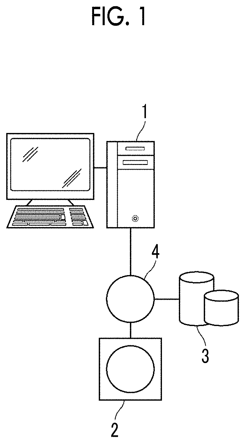
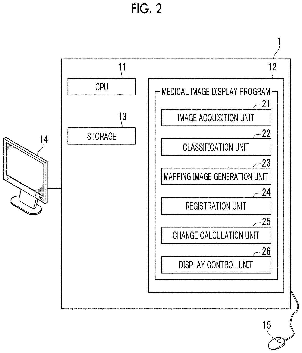
[0037]Hereinafter, an embodiment of the invention will be described with reference to the accompanying diagrams. FIG. 1 is a hardware configuration diagram showing the outline of a diagnostic support system to which a medical image display device according to an embodiment of the invention is applied. As shown in FIG. 1, in the diagnostic support system, a medical image display device 1 according to the present embodiment, a three-dimensional image capturing apparatus 2, and an image storage server 3 are communicably connected to each other through a network 4.

[0038]The three-dimensional image capturing apparatus 2 is an apparatus that generates a three-dimensional image showing a part, which is a part to be examined of a subject, by imaging the part. Specifically, the three-dimensional image capturing apparatus 2 is a CT apparatus, an MRI apparatus, a positron emission tomography (PET) apparatus, or the like. The three-dimensional image generated by the three-dimensional image capt...


PUM


Abstract

A classification unit classifies a lung region, which is included in each of two three-dimensional images having different imaging times for the same subject, into a plurality of types of case regions. A mapping image generation unit generates a plurality of mapping images corresponding to the three-dimensional images by labeling each of the classified case regions. A change calculation unit calculates the center-of-gravity position of each case region for case regions at corresponding positions in the mapping images, and calculates the movement amount and the movement direction of the center-of-gravity position between the mapping images as a change of each case region. A display control unit displays information regarding the change on a display.

Description

CROSS REFERENCE TO RELATED APPLICATIONS[0001]The present application claims priority under 35 U.S.C. § 119 to Japanese Patent Application No. 2017-077361 filed on Apr. 10, 2017. The above application is hereby expressly incorporated by reference, in its entirety, into the present application.BACKGROUNDTechnical Field[0002]The present invention relates to a medical image display device, method, and program for identifying and displaying a plurality of types of case regions included in a medical image.Related Art[0003]Image diagnosis using medical images obtained by capturing a radiographic image and an ultrasound image of a patient, who is a subject, has been performed. In recent years, due to advances in medical apparatuses such as computed tomography (CT) apparatuses and magnetic resonance imaging (MRI) apparatuses, high-quality three-dimensional images with high resolution are used for image diagnosis.[0004]Incidentally, interstitial pneumonia is known as a disease of the lung. A ...

Claims


Application Information

Patent Timeline
Patent Type & Authority Patents(United States)
IPC IPC(8): A61B5/00G06T19/00G06T7/00G06T11/00A61B8/08G06T15/08G06T7/11G06K9/62G06V10/764
CPCA61B5/748A61B8/08G06K9/6273G06T7/0012G06T7/0016G06T11/00G06T15/08G06T19/00A61B5/7425A61B8/483G06T7/11G06T2200/24G06T2207/10072G06T2207/20084G06T2207/30061G06T2210/41G06V10/82G06V10/764G06F18/2414
Inventor OSAWA, AKIRA
Owner FUJIFILM CORP
Why Eureka
Try Eureka