
Method of obtaining a population of cells

a cell population and cell technology, applied in the field of cell population obtaining, can solve the problems of limited current state of the art, damage, and inability to easily access the collection site (the bone marrow), and achieve the effect of convenient collection

Active Publication Date: 2019-04-30
RIGENERAND
View PDF8 Cites 0 Cited by
  • Summary
  • Abstract
  • Description
  • Claims
  • Application Information

AI Technical Summary

Benefits of technology

The invention provides a method for obtaining a population of cells that can be used for therapeutic purposes. The method allows for the collection of small amounts of tissue from a variety of patients through minimally invasive surgery, which reduces pain and discomfort for the patient. The collected tissue can also be easily extended to a larger number of individuals, making it a useful treatment tool. Overall, the invention provides a way to obtain and develop therapeutic cells from small amounts of accessible tissue.

Problems solved by technology

This is required because of the poor presence of SP in the original bone marrow tissue, and may be a limitation.
Furthermore, the collection site (the bone marrow) may not be easily accessible and be damaged due to the presence of neoplastic cells or simultaneous pharmacological treatments.
The present state of the art is limited by the large amounts of subcutaneous AT that must be collected to obtain an adequate number of SP.
Therefore, this method in the prior art is only applicable to a limited number of patients.
Moreover, invasive surgical procedures are required, involving the hazards of any surgery, particularly fat embolism.
Further, AT collections always require a general anesthesia of the patient, with the related requited care and potential hazards.

Method used


Image

  • Method of obtaining a population of cells
  • Method of obtaining a population of cells
  • Method of obtaining a population of cells

Examples


example 1

Production of a Population with Desired Characteristics

[0069]An AT sample was collected from the subcutaneous facial area of a healthy 46-year old female donor by Coleman liposuction. Alternate methods may be used for collection.

[0070]The AT fragments were washed three times with a saline solution known as Dulbecco-phosphate buffer solution (D-PBS) added with antibiotic (1 U / mL penicillin, 1 mg / mL streptomycin and 2.5 mg / mL amphotericin B), for an overall time of 15 minutes.

[0071]The last washing step, which was designed to yield a sample with no liquid component, was carried out using a filter (100 μm cell strainer).

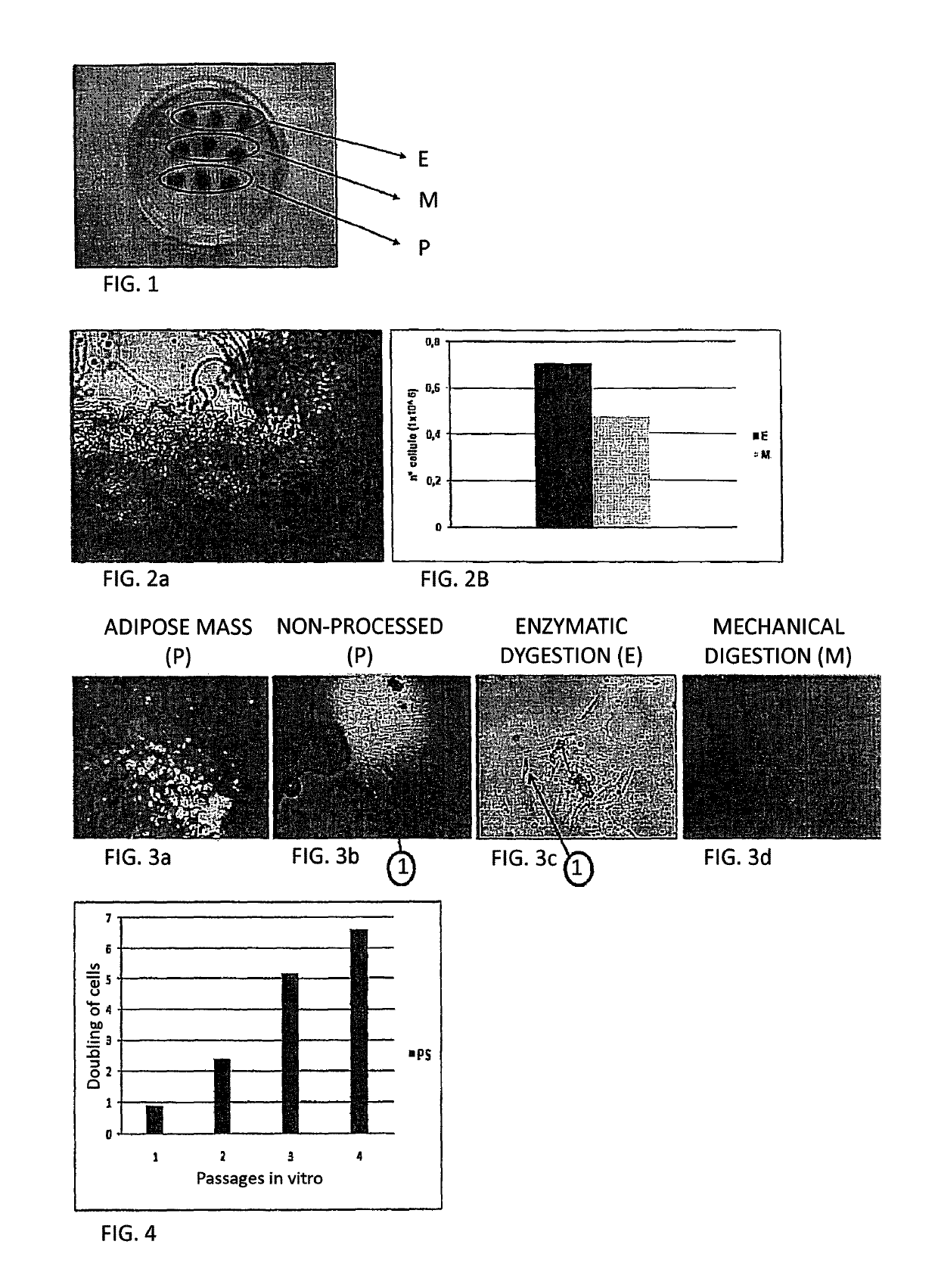
[0072]The AT fragments were transferred into a sterile container (Petri dish) and divided into nine parts, each having a volume of about 0.025 mL. The latter, as shown in FIG. 1, underwent three different procedures in triplicate: three parts (sample E) underwent the same enzyme digestion process, three parts (sample M) underwent the same mechanical digestion process an...

example 2

Immunophenotyping Evaluation

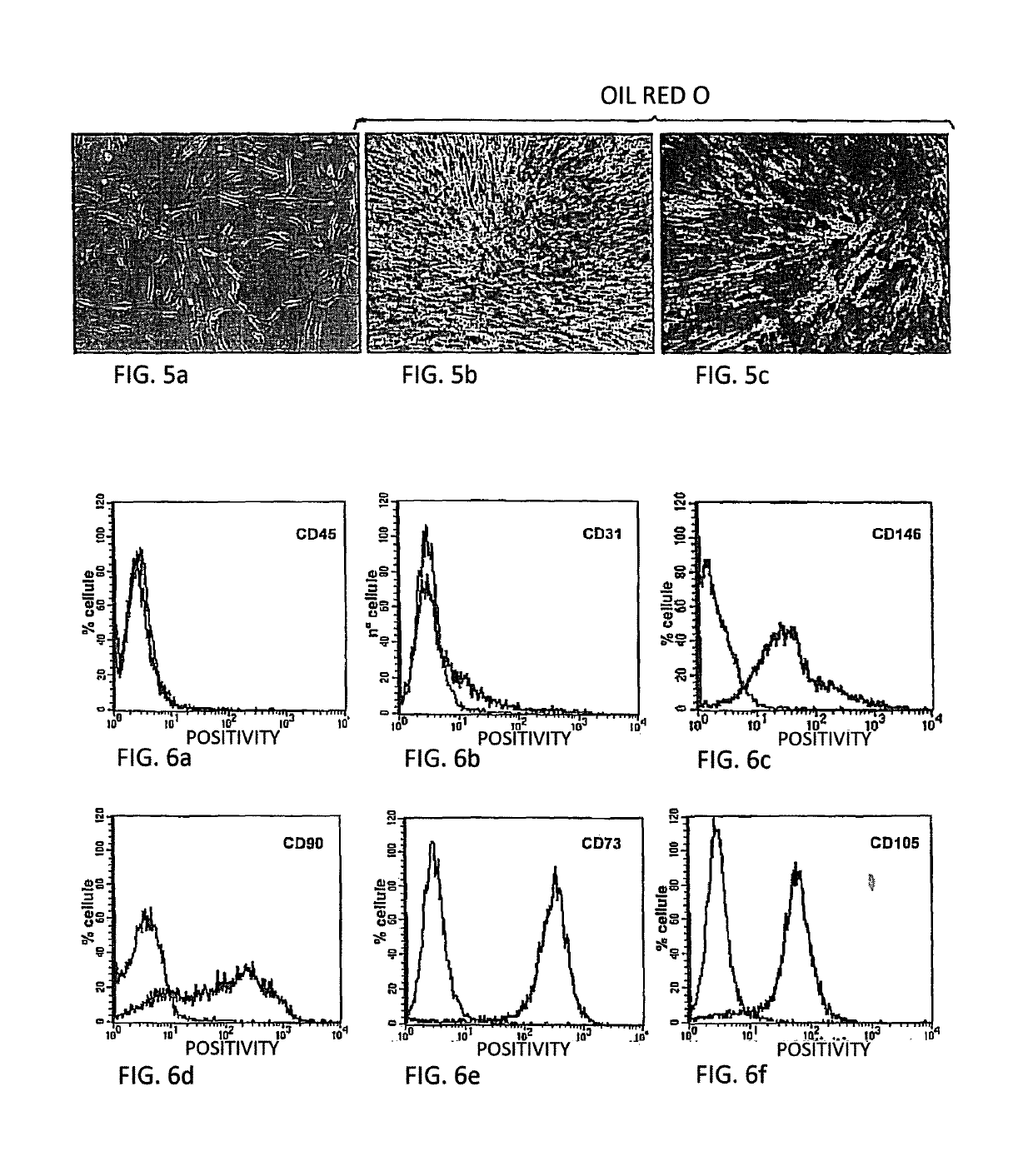
[0094]A check was first made for the presence of any contaminating population, such as endothelial cells and immune system cells, by searching for their respective CD31 and CD45 markers. The SP were found to be negative for CD45 (FIG. 6a) and very weakly positive for CD31 (FIG. 6b).

[0095]At the same time, the SP were found to be positive for progenitor antigens, such as CD90 (FIG. 6d), CD105 (FIG. 6f) and CD73 (FIG. 6e) and to a lesser extent for CD146 (FIG. 6c) as a typical stromal pericyte marker.

example 3

Differentiation Assays

[0096]Osteogenic, adipogenic and chondrogenic and differentiation of the SP were performed after phenotyping, at passage 4.

[0097]Osteogenic Differentiation.

[0098]The cells were seeded at a density of 10,000 cells / cm2. After full growth (typically after 2-4 days), they were induced osteogenic differentiation, with one sample being preserved as control.

[0099]Bone induction was obtained using an appropriate medium, composed of: basal medium (DMEM with 10% fetal calf serum—FCS) dexamethasone, L-ascorbic-2-phosphate acid and β-glycerophosphate.

[0100]Such basal medium was maintained for one week, and replaced every 2-3 days. On the seventh day, or day 7, bone morphogenetic protein-2 was added to the medium. The cells were maintained with the differentiating medium for seven more days, and such medium was replaced every two-three days.

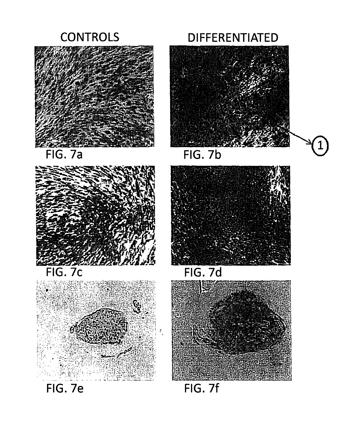
[0101]On the fourteenth day, the differentiation result was assessed by histological assay (Alizarin Red staining). In this assay, the ...


PUM

PropertyMeasurementUnit
temperatureaaaaaaaaaa
volumesaaaaaaaaaa

Abstract

A method of obtaining stromal progenitor cells (SPC) from subcutaneous adipose tissue by incubation of a very small volume of the subcutaneous adipose tissue in an enzyme solution produces SPC that are usable in medical applications based on autologous SPC even on individuals having a body mass index lower than 18.5.

Description

FIELD OF THE INVENTION[0001]The present invention relates to a method of obtaining a population of cells, particularly a population of stromal progenitor cells from a quantity of white adipose tissue collected from a living being.BACKGROUND ART[0002]Stromal progenitors (SP) are cell populations that are capable of proliferation and differentiation, and also provide support for surrounding tissues and cells.[0003]SP are rare cell elements located in the tissues of living beings, which are designed to perpetuate their function by turnover of damaged and / or senescent cells.[0004]SP were initially found in high-turnover tissues, such as the hematopoietic and epithelial system, but can be found, although less frequently, in tissues and organs having little or no regenerative capacity, such as the central nervous system.[0005]Originally, bone marrow studies started more than thirty years ago were able to identify certain SP, which will be referred to hereinafter as mesenchymal SP, which a...

Claims


Application Information

Patent Timeline
Patent Type & Authority Patents(United States)
IPC IPC(8): C12N5/0775
CPCC12N5/0667C12N2509/00
Inventor DOMINICI, MASSIMOCAFARELLI, LUIGIVERONESI, ELENAPICCINNO, MARIA SERENAPAOLUCCI, PAOLODE SANTIS, GIORGIOCONTE
Owner RIGENERAND
Learn More