
System and method for assessing perfusion in an anatomical structure

a technology of anatomical structure and hemodynamics, applied in the field of system for measuring and assessing hemodynamics in anatomical structure, can solve the problems of frequent postsurgical complications in connection with anastomosis in the gastrointestinal tract, tissue damage, and anastomotic leakage, so as to reduce post-surgical complications, reduce the risk of leakage, and prolong the hospital stay.

Active Publication Date: 2019-04-30
PERFUSION TECH IVS
View PDF8 Cites 0 Cited by
  • Summary
  • Abstract
  • Description
  • Claims
  • Application Information

AI Technical Summary

Benefits of technology

[0024]A reliable procedure to assess perfusion in the gastrointestinal tract, in particular the wall of the gastrointestinal tract is therefore needed, more specifically before, during and after examination and / or surgery. In connection with bowel surgery where bowel resection, and possibly anastomosis, is needed a more reliable procedure is needed to reduce post-surgical complications. Today the surgeon is left with subjective parameters to assess perfusion, for example palpable pulse, color of tissue and active bleeding. Most advantageously the surgeon should be able to objectively quantify the perfusion intraoperatively such that decisions can be evaluated and revised during surgery. Assessment of the perfusion of, for example, an anastomosis would therefore be of great interest because it would allow the surgeon to intervene in appropriate situations, thereby possibly reducing the risk of leakage and prolonged hospital stay which increases the healthcare costs.
[0025]One solution could be the application of fluorescence imaging using a fluorescent dye injected in the blood of the patient. Fluorescence imaging in the form of Laser Fluorescence Angiography (LFA) has a proved correlation to perfusion [1]. However, an efficient utilization of fluorescence imaging requires signal processing in order to quantify the perfusion. It would be advantageous if tools were provided for the surgeon to objectively evaluate perfusion before, during and / or after surgery, for example before resection, before anastomosis and / or after anastomosis. The purpose of the present disclosure is therefore also to contribute to the improvement within diagnosis and treatment procedures of the gastrointestinal tract, in particular intestinal anastomosis, e.g. in connection with colorectal cancer treatment.
[0042]The methods disclosed herein are advantageously computer implemented to provide for fast and reliable image processing and data analysis, possibly in real time or at least near real time such that they can be used intraoperative.
[0043]The presently disclosed method for obtaining a measure of hemodynamics may in a further embodiment contribute to an assessment of a patient's risk of postsurgical complications and thereby for example help the surgeon to make an informed choice of an appropriate follow-up program following examination or surgery personalized to each patient. One way to do this could be to combine the calculated perfusion parameter(s) with various relevant patient specific data such as age, gender, comorbidities, type of surgery etc. in a multivariate analysis, providing the surgeon with an automatically weighted risk score for each patient already during the surgery. Such an intelligent risk score can contribute to the surgeons' surgical decisions during any operation on the gastrointestinal tract, where a measurement of perfusion could be of interest.
[0044]By storing each patient's data from the multivariate analysis in an online database, preferably in fully anonymized form, this growing database can then be used to continuously update and refine the analysis itself. Resulting in an ever improving analysis and a decision support system based on each patient's personalized risk profile. I.e. what can be provided is a decision support system that aids the surgeon both during the operation to help reduce the risk of postsurgical complications, and after the operation when selecting the appropriate follow-up program.

Problems solved by technology

Postsurgical complications in connection with anastomosis in the gastrointestinal tract are unfortunately frequent, often due to insufficient perfusion (capillary blood supply) at the anastomosis, i.e. the joining of the two parts of the tract.
Insufficient perfusion may cause anastomotic leakage, which is a serious and frequent complication, for example in connection with colorectal surgery where more than 10% of the procedures result in complications.
Risk factors associated with leakage include tension of anastomosis, tissue damage and in particular reduced blood perfusion.

Method used


Image

  • System and method for assessing perfusion in an anatomical structure
  • System and method for assessing perfusion in an anatomical structure
  • System and method for assessing perfusion in an anatomical structure

Examples


examples

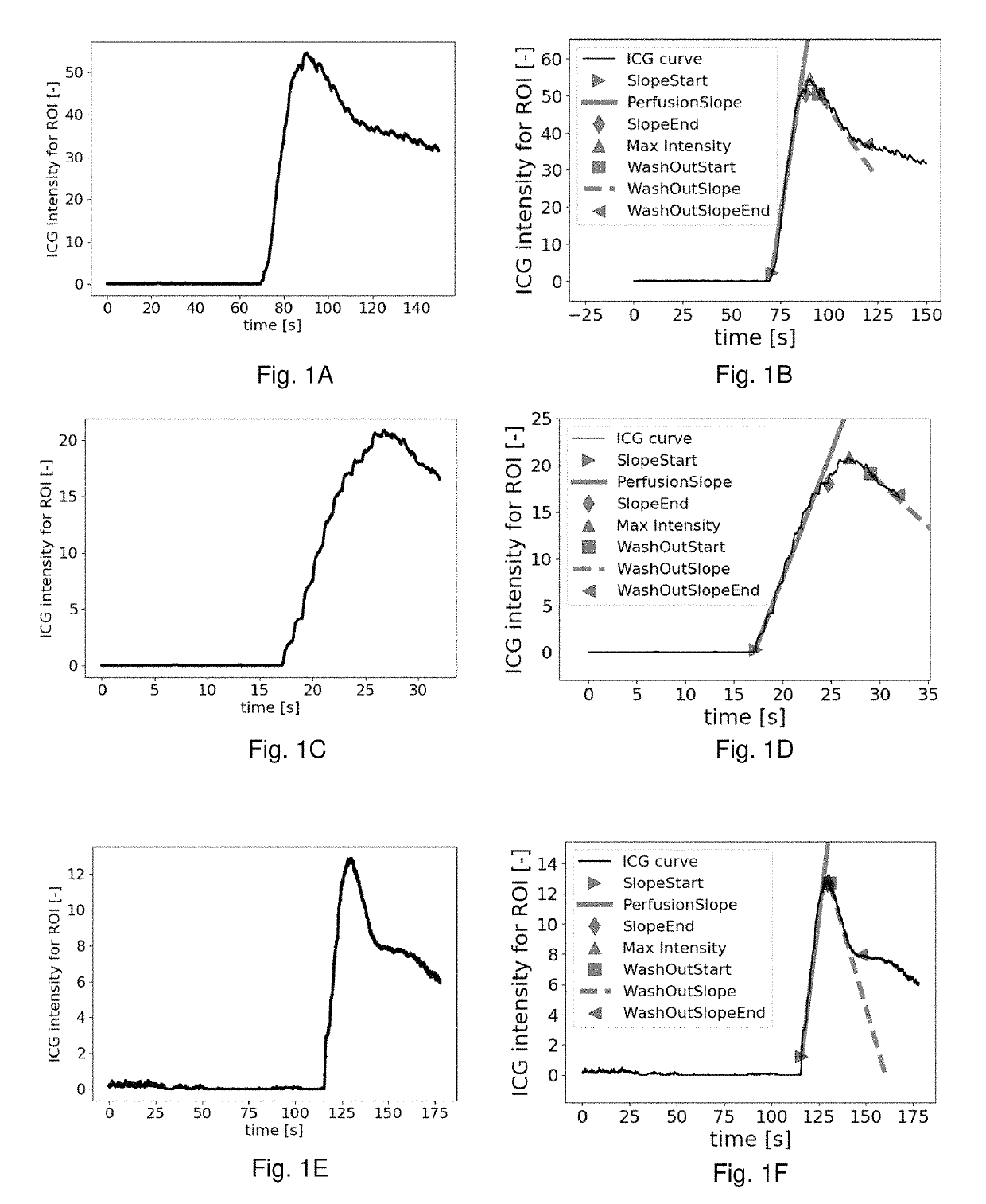
[0110]FIGS. 1A, 1C and 1E show examples of intensity curves acquired from tissue after a bolus of ICG has been provided to a subject, e.g. from a region of interest in a video sequence. The same kind of data could be obtained if another contrast agent was used. The intensity is substantially zero until a steep rise in intensity indicates the passage of ICG molecules in the imaged tissue, the ICG molecules being excited to fluoresce. The peak in intensity is followed by the gradual washout of the ICG molecules. The intensity is indicated with arbitrary units. FIGS. 1B, 1D, and 1F show the corresponding intensity curves where the hemodynamic parameters perfusion slope, slope start, slope end max intensity, washout slope, washout start and washout slope end have been calculated and are indicated in the graphs.

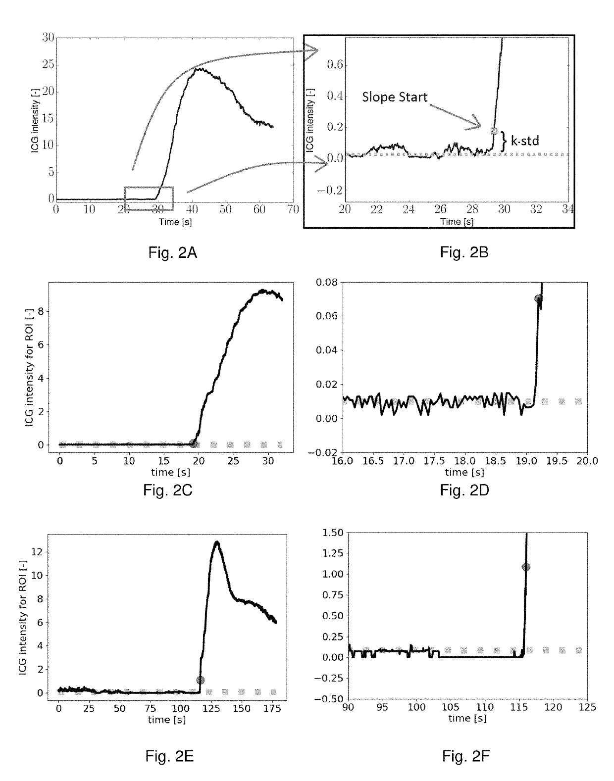
[0111]FIGS. 2A-2F show three examples illustrating the herein disclosed approach of determining the point in time where the perfusion slope starts, i.e. slope start. FIGS. 2B, 2D ...


PUM


Abstract

A system is provided for measuring and assessing hemodynamics in an anatomical structure of a subject, along with a method for image processing hemodynamics in at least a part of an anatomical structure in video images acquired from a subject. The system and method relates to measuring and assessing hemodynamics in, around and near the surface, in particular the gastrointestinal wall, of the gastrointestinal tract of a subject. A method for image processing hemodynamics in at least a part of an anatomical structure in video images acquired from a subject may performing image analysis of at least one video sequence acquired after a fluorescent contrast agent has been supplied to the subject, calculating intensity values in one or more regions of interest based on the image analysis, and determining the perfusion slope of the flow of the fluorescent contrast agent through at least one of said regions of interest.

Description

CROSS REFERENCE TO RELATED APPLICATIONS[0001]This application claims priority from European Application No. 16203188.4 filed Dec. 9, 2016, the entire content of which is incorporated herein by reference.BACKGROUND OF INVENTION[0002]The present disclosure relates to a system for measuring and assessing hemodynamics in an anatomical structure of a subject, and a method for image processing hemodynamics in at least a part of an anatomical structure in video images acquired from a subject. In particular the present disclosure relates to measuring and assessing hemodynamics in, around and near the surface, in particular the gastrointestinal wall, of the gastrointestinal tract of a subject.[0003]Complications related to the gastrointestinal tract are often related to local hemodynamics. I.e. a change in the normal hemodynamic conditions may be an indicator of a complication. Perfusion assessment of the gastrointestinal tract, in particular in and near the surface of the gastrointestinal t...

Claims


Application Information

Patent Timeline
Patent Type & Authority Patents(United States)
IPC IPC(8): A61B5/0275A61B5/026A61B5/00A61B17/11A61B17/00A61B5/11
CPCA61B5/0275A61B5/0261A61B5/4255A61B5/4552A61B5/7225A61B5/748A61B17/1114A61B5/0071A61B2576/00A61B5/0088A61B5/1128A61B5/4233A61B5/4238A61B5/4848A61B5/7264A61B2017/00818
Inventor MADEN, MADS HOLST AAGAARDLUND, MORTEN TOFT
Owner PERFUSION TECH IVS
Learn More