Vessel segmentation

a technology of x-ray and segmentation, applied in the field of x-ray segmentation, can solve the problems of adding x-ray dose, and achieve the effect of reducing x-ray dose, improving and facilitating

Active Publication Date: 2018-10-02
KONINKLJIJKE PHILIPS NV
View PDF15 Cites 1 Cited by
  • Summary
  • Abstract
  • Description
  • Claims
  • Application Information

AI Technical Summary

Benefits of technology

[0004]There may thus be a need to provide an improved and facilitated way of providing vascular flow and surrogate vessel geometry information with reduced X-ray dose.
[0007]This provides the advantage that although only a 2D sequence of images is provided, depth information is provided nevertheless by taking into account the time factor related to the arrival of a contrast agent, for example, which then can be used for the segmentation data calculation. The sequence comprises only one 2D image for each point in time, thus meaning a minimum image dose exposure for the patient. The surrogate vessel geometry information is thus provided based on image acquisition with reduced X-ray dose, and only one geometry configuration, also facilitating the acquisition itself. Furthermore, since the segmentation data is derived from the same angiogram used for further purposes, there are no differences due to patient movement or vascular changes such as vasodilation that could occur when an extra acquisition is used for segmentation.
[0009]This provides the advantage that the vessel confidence index provides further detailed, and thus improved, information relating to the segmentation.
[0011]This provides the advantage in that the provision of the segmentation outline further improves the visible depth appearance, or in other words, further supports to provide three-dimensional information although the images, on which the presented displayed image is based on, only comprise 2D images and not 3D images.
[0014]For example, the object is a patient under observation, for example in a hospital. The provision of the injection device and a data and signal connection to the X-ray image processing device allows regulating the amount of contrast agent to be minimal.
[0030]According to an aspect of the present invention, the analysis of contrast arrival time is used for segmenting the image sequence; and the segmentation may be used in a 2D flow calculation, for example automatically. Thereby it is possible to calculate the connectivity per pixel, and therefore the invention allows separating and identifying false connections of crossing overlapping vessels or vessels touching each other in the X-ray image. The connectivity information can then be used as a mask in the flow extraction algorithm. The computation of a contrast arrival time map using an analysis of each pixel grey level along the time series, leads to an estimation of the arrival time of the contrast agent within each pixel of the whole image. In addition with a vessel confidence map, the quality of the arrival time map is further improved, leading to improved visual impression of the information for the user. This can then be used for segmentation, wherein the connectivity based on arrival time maps permits to distinguish different vessels that are superposed by projection. The identification of disconnected flow parts allows separately processing each non-overlapping segment and discarding vessel overlapping parts from the processing. It further allows identifying where specific processing techniques have to be used to extract flow field in the overlapping areas. The term “flow” relates to contrast displacement in the sequence, and thus to blood velocity.

Problems solved by technology

However, this means an additional X-ray dose and requires either that the X-ray system be moved, in which case the segmentation data cannot be obtained at the same time as the flow acquisition, or it requires the use of multiple X-ray imagers.

Method used


Image

  • Vessel segmentation
  • Vessel segmentation
  • Vessel segmentation

Examples


Embodiment Construction

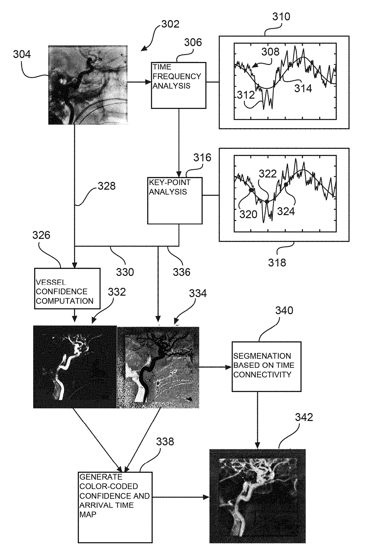
[0042]FIG. 1 shows an X-ray image processing device 10, comprising an interface unit 12 and a data processing unit 14. The interface unit 12 is configured to provide a sequence of time series angiographic 2D images of a vascular structure obtained after a contrast agent injection. For example, the series of 2D images is provided by a (not shown) data base. In another example, the 2D images are provided by an X-ray imaging system (see also further below). The data processing unit 14 is configured to determine an arrival time index of a predetermined characteristic related to the contrast agent injection for each of a plurality of determined pixels along the time series. The data processing unit 14 is also configured to compute a connectivity index for each of the plurality of the determined pixels based on the arrival time index. The data processing unit is further configured to generate segmentation data of the vascular structure from the plurality of the determined pixels, wherein ...


PUM


Abstract

An X-ray image processing device for providing segmentation information with reduced X-ray dose that includes an interface unit, and a data processing unit. The interface unit is configured to provide a sequence of time series angiographic 2D images of a vascular structure obtained after a contrast agent injection. The data processing unit is configured to determine an arrival time index of a predetermined characteristic related to the contrast agent injection for each of a plurality of determined pixels along the time series, and to compute a connectivity index for each of the plurality of the determined pixels based on the arrival time index. The data processing unit is configured to generate and provide segmentation data of the vascular structure from the plurality of the determined pixels, wherein the segmentation data is based on the connectivity index of the pixels.

Description

CROSS-REFERENCE TO PRIOR APPLICATIONS[0001]This application is the U.S. National Phase application under 35 U.S.C. § 371 of International Application No. PCT / IB2014 / 060371, filed on Apr. 2, 2014, which claims the benefit of European Patent Application No. 13305426.2, filed on Apr. 3, 2013. These applications are hereby incorporated by reference herein.FIELD OF THE INVENTION[0002]The present invention relates to vessel segmentation, and relates in particular to a method for providing segmentation information, to an X-ray image processing device, and to an X-ray imaging system, as well as to a computer program element and a computer readable medium.BACKGROUND OF THE INVENTION[0003]In medical X-ray imaging, segmentation of vessels is used for providing enhanced information about a vessel structure, for example. Such information can be used for assessing a health state of a patient or also for planning of a necessary intervention, but also for live guidance during an intervention, and a...

Claims


Application Information

Patent Timeline
Patent Type & Authority Patents(United States)
IPC IPC(8): G06T7/20A61B6/00G06T7/187G06T7/174G06T7/11
CPCG06T7/20A61B6/481A61B6/504A61B6/52G06T7/11G06T7/187G06T7/174G06T2207/30104G06T2207/10016G06T2207/10116G06T2207/30101
Inventor STAWIASKI, JEANBONNEFOUS, ODILERUIJTERS, DANIEL SIMON ANNAVAN NIJNATTEN, FRED SIMON BEREND
Owner KONINKLJIJKE PHILIPS NV
Why Eureka
Try Eureka