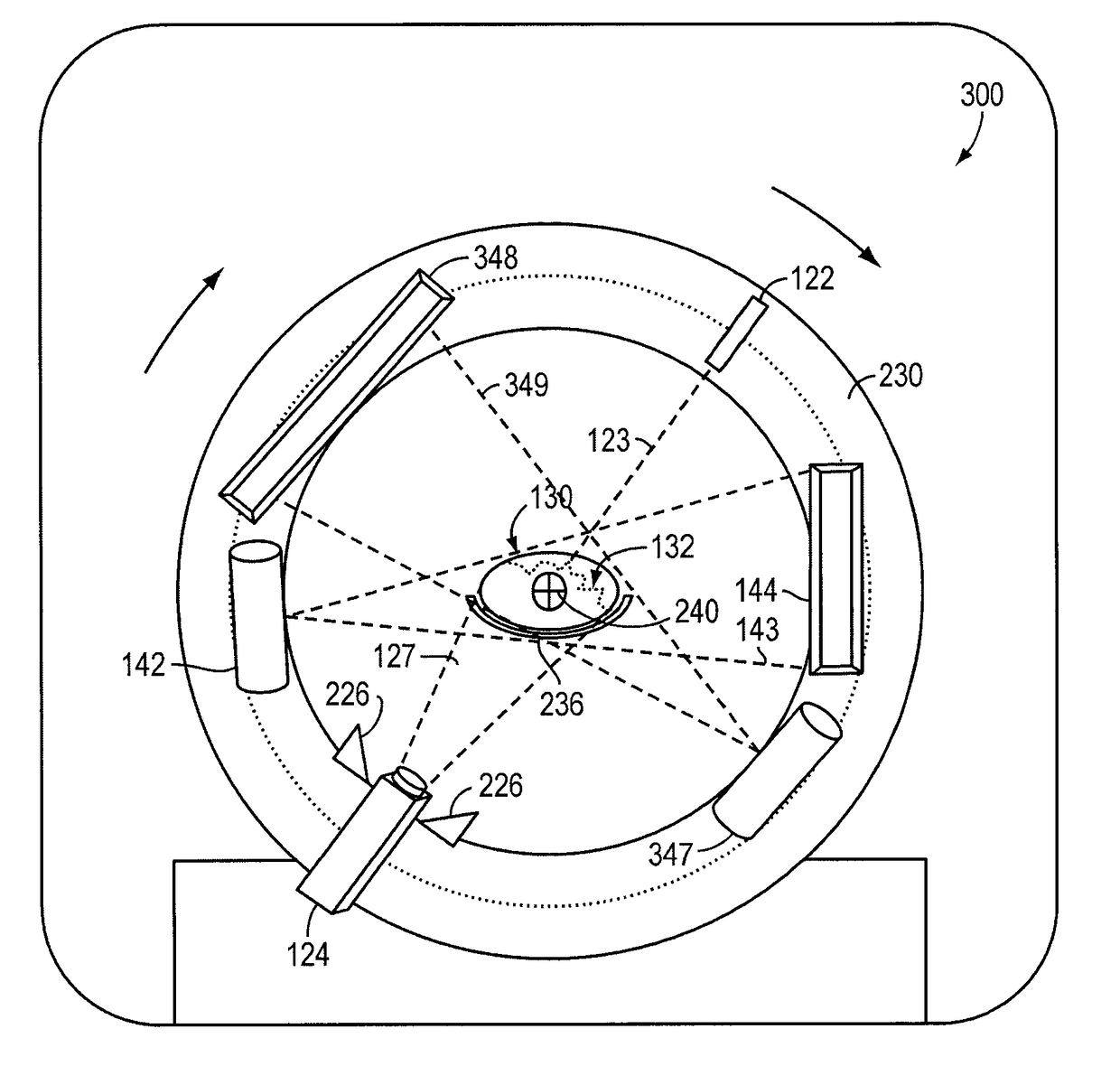
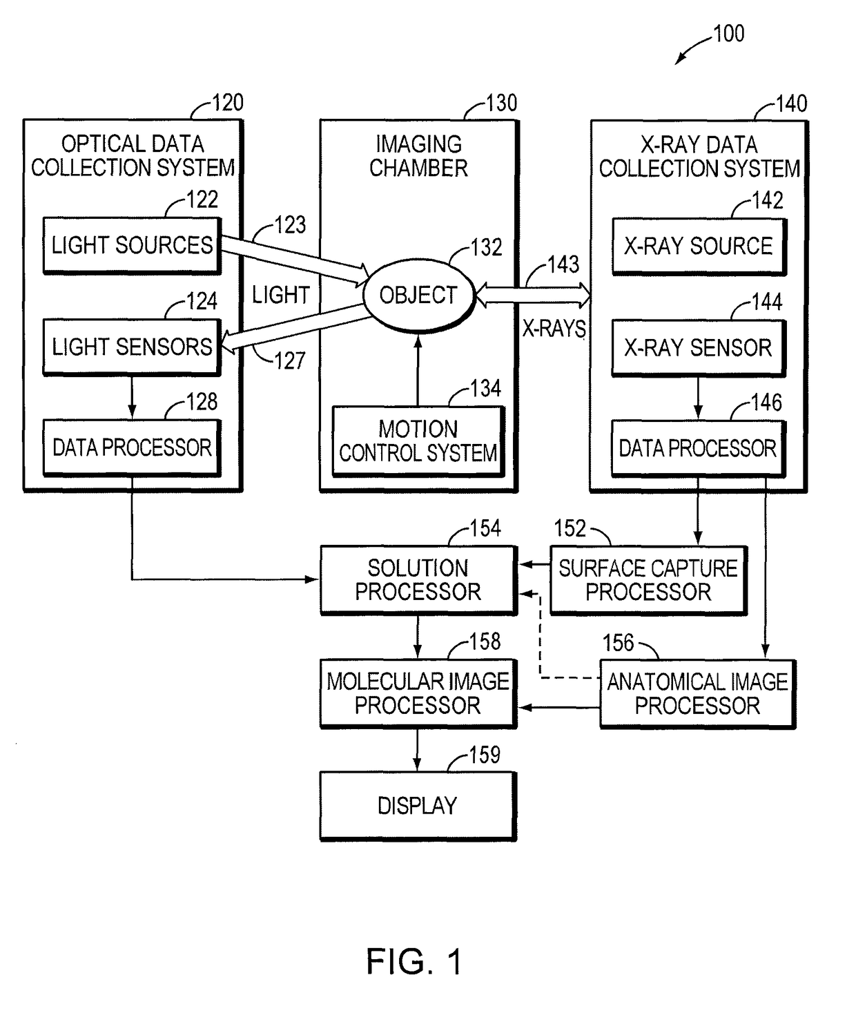
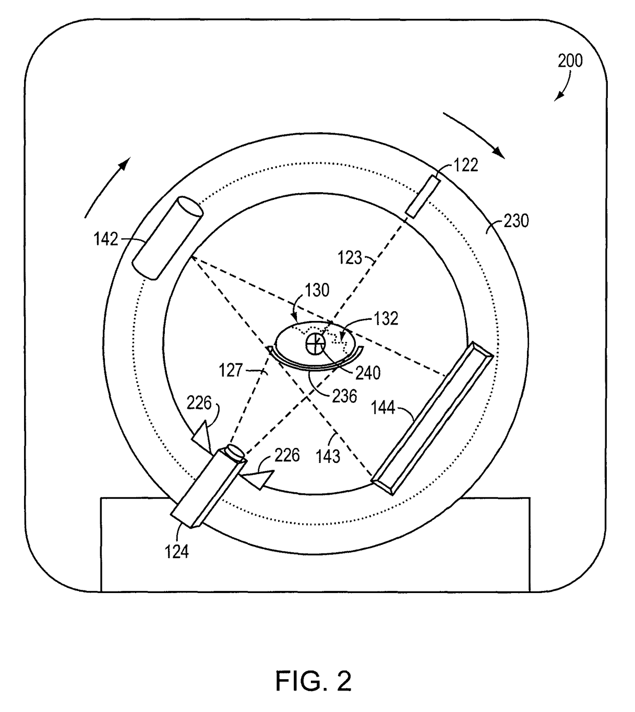
Combined x-ray and optical tomographic imaging system
a combined x-ray and optical tomographic technology, applied in tomography, applications, instruments, etc., can solve the problems of insufficient information contained in the signal received by a single energy sensor (e.g., at one angle), inability to detect, distinguish, and existing x-ray cat systems cannot provide functional (or, “molecular”) information about a subject or disease state at the cellular or molecular level, and achieve accurate functional information
- Summary
- Abstract
- Description
- Claims
- Application Information
AI Technical Summary
Benefits of technology
Problems solved by technology
Method used
Image

Examples
Embodiment Construction
[0086]It is contemplated that methods, systems, and processes described herein encompass variations and adaptations developed using information from the embodiments described herein.
[0087]Throughout the description, where systems and compositions are described as having, including, or comprising specific components, or where processes and methods are described as having, including, or comprising specific steps, it is contemplated that, additionally, there are systems and compositions of the present invention that consist essentially of, or consist of, the recited components, and that there are processes and methods of the present invention that consist essentially of, or consist of, the recited processing steps.
[0088]The mention herein of any publication, for example, in the Background section, is not an admission that the publication serves as prior art with respect to any of the claims presented herein. The Background section is presented for purposes of clarity and is not meant a...
PUM
 Abstract
Description
Claims
Application Information
 - R&D
- Intellectual Property
- Life Sciences
- Materials
- Tech Scout
- Unparalleled Data Quality
- Higher Quality Content
- 60% Fewer Hallucinations
Browse by: Latest US Patents, China's latest patents, Technical Efficacy Thesaurus, Application Domain, Technology Topic, Popular Technical Reports.
© 2025 PatSnap. All rights reserved.Legal|Privacy policy|Modern Slavery Act Transparency Statement|Sitemap|About US| Contact US: help@patsnap.com