
Consistent sequential ultrasound acquisitions for intra-cranial monitoring

a technology of intra-cranial monitoring and sequential ultrasound, which is applied in the field of positioning of medical imaging probes, can solve the problems of common delay of about one hour to perform ct scans, inability to move patients from the bed to the ct room for repeated follow-up, and inability to perform repeated ct examinations. to achieve the effect of quick ascertaining presence and monitoring

Active Publication Date: 2018-07-31
KONINKLJIJKE PHILIPS NV
View PDF27 Cites 3 Cited by
  • Summary
  • Abstract
  • Description
  • Claims
  • Application Information

AI Technical Summary

Benefits of technology

[0016]No device presently exists to quickly ascertain presence and monitoring of intra-cranial hemorrhage that can be used in an ER or pre-hospital setting without significant user expertise.

Problems solved by technology

While CT can be used for the initial assessment of trauma and stroke victims, repeated CT examinations are not advisable due to the high radiation dose.
Also, it is impractical to move the patient from the bed to the CT room for repeated follow-ups.
In many centers, a delay of about one hour to performing a CT scan is common.
Transcranial ultrasound scans, however, are difficult to perform and require considerable expertise.
This is because changes in the probe position and / or angle lead to drastically different acoustic windows due to the uneven nature of the skull, leading to different transmit amplitudes and backscattering properties.

Method used


Image

  • Consistent sequential ultrasound acquisitions for intra-cranial monitoring
  • Consistent sequential ultrasound acquisitions for intra-cranial monitoring
  • Consistent sequential ultrasound acquisitions for intra-cranial monitoring

Examples


Embodiment Construction

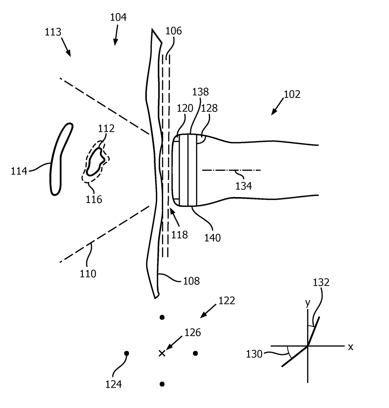
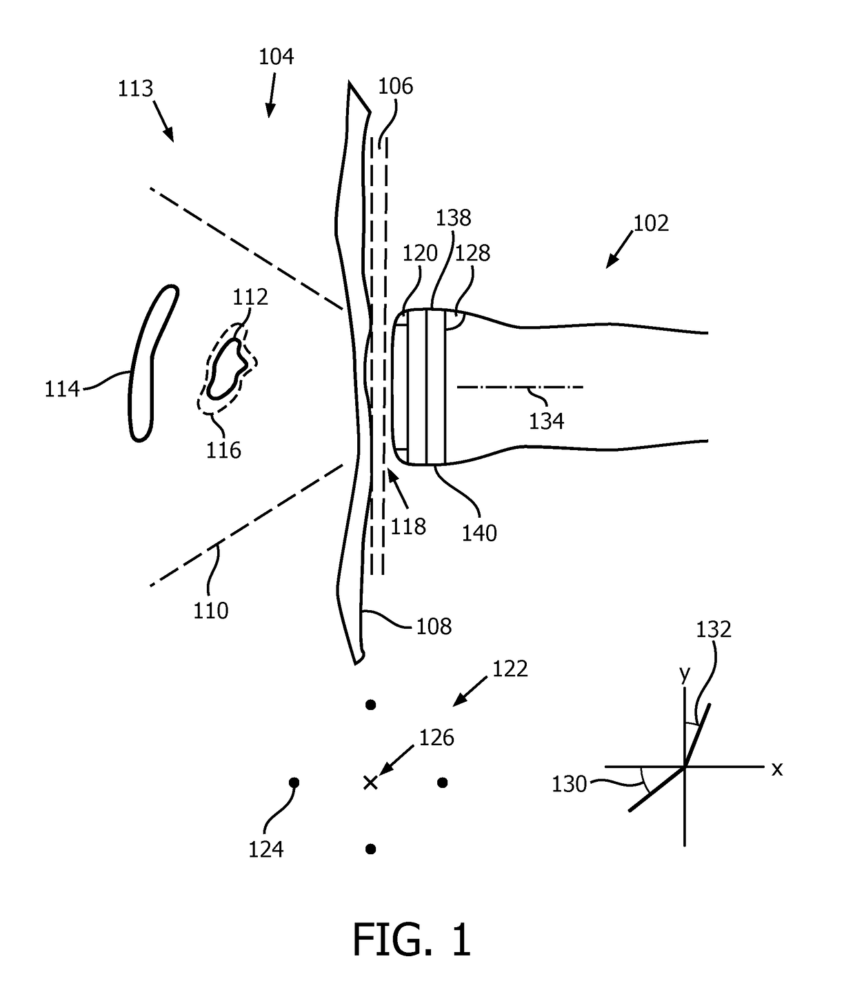
[0048]FIG. 1 depicts an exemplary probe 102 for consistent sequential ultrasound acquisitions for intra-cranial monitoring of a medical subject 104, such as a human patient or animal.

[0049]It will be assumed, in the description that follows, that this single probe 102 is used for all the acquisitions. However, a medical imaging device could feature one or more other probes, any one of which can be applied to the subject 104 for a given acquisition. In this disclosure, any of the probes, if there are more than one, could be characterized as “a probe said medical imaging device comprises.” Otherwise, if there is a single probe, that probe is “a probe said medical imaging device comprises.”

[0050]The probe 102 is applied to skin 106 of the subject 104 in the temple area of the head. The probe 102 is therefore pressed against an underlying, bony structure, a temporal bone 108. Due to the uneven nature of the temporal bone 108, changes in the probe position and / or angle lead to drasticall...


PUM


Abstract

A medical imaging probe (102) for contact with an imaging subject includes an indicium placement apparatus for, while the probe is in contact, selectively performing an instance of marking the subject so as to record a position of the probe. The device may further include a feedback module for determining whether an orientation, with respect to a the mark, that currently exists for a medical imaging probe of the device meets a criterion of proximity to a predetermined orientation. Responsive to the determination that the criterion is met, a quantitative evaluation may be made automatically and without need for user intervention, via live imaging via the probe, of a lesion that was, prior to the determination, specifically identified for the evaluation. Change, such as growth (116), in the lesion, like a brain lesion, may thereby be tracked over consistent sequential imaging acquisitions, such as through ultrasound.

Description

CROSS-REFERENCE TO PRIOR APPLICATIONS[0001]This application is the U.S. National Phase application under 35 U.S.C. § 371 of International Application No. PCT / IB2014 / 059232, filed on Feb. 25, 2014, which claims the benefit of U.S. Provisional Patent Application No. 61 / 772,717, filed on Mar. 5 2013. These applications are hereby incorporated by reference herein.FIELD OF THE INVENTION[0002]The present invention relates to a position of a medical imaging probe and, more particularly, to the position while the probe is in contact with an imaging subject.BACKGROUND OF THE INVENTION[0003]According to the Centers for Disease Control and Prevention (CDC), trauma accounts for 42 million emergency department visits and 2 million hospital admissions across the USA every year. In the USA, trauma accounts for 38.4 deaths per 100,000 of population. Trauma is especially a leading cause of death in the young population, and accounts for 30% of all life years lost in the USA (compared to 16% for canc...

Claims


Application Information

Patent Timeline
Patent Type & Authority Patents(United States)
IPC IPC(8): A61B8/00A61B5/00A61B8/14A61B8/08A61B90/00
CPCA61B5/6842A61B8/0808A61B8/14A61B8/42A61B8/54A61B8/46A61B8/5223A61B8/4245A61B2090/395
Inventor RAJU, BALASUNDAR IYYAVUSHI, WILLIAM TAOVIGNON, FRANCOIS GUY GERARD MARIE
Owner KONINKLJIJKE PHILIPS NV
Learn More