Quantum point marker sandwich immunodetection method and its diagnosis kit
A technology of sandwich immunity and quantum dots, applied in biological testing, measuring devices, material inspection products, etc., can solve the problems of unstable color development, long detection cycle, cumbersome operation, etc., and achieve simple and easy operation, strong The effect of resistance
- Summary
- Abstract
- Description
- Claims
- Application Information
AI Technical Summary
Problems solved by technology
Method used
Image
Examples
Embodiment 1
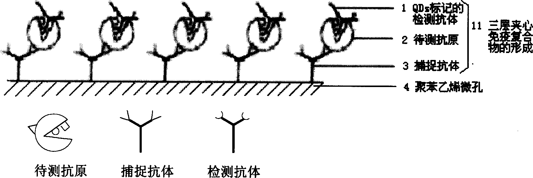
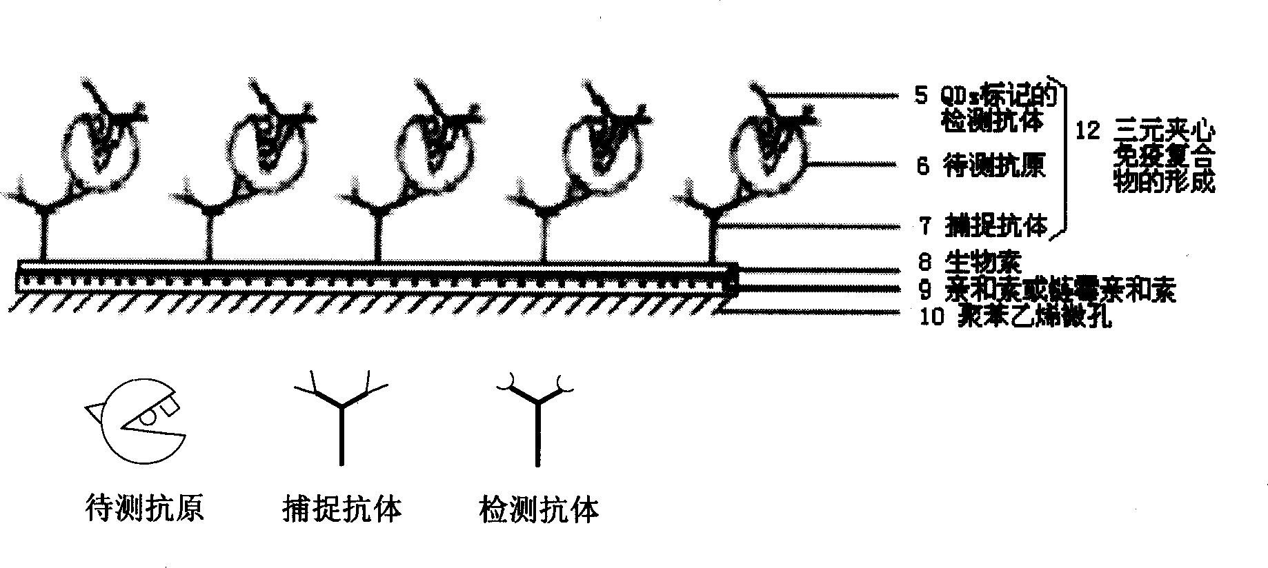
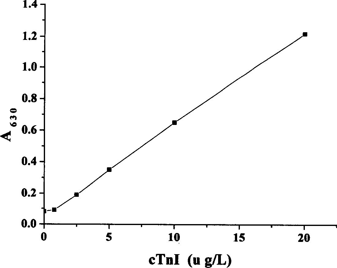
[0039] Quantum dot-labeled sandwich immunoassay of cardiac troponin I (cTnI)
[0040] (1) Anti-cTnI monoclonal antibody was labeled with red light-emitting QD630 to prepare detection antibody
[0041]Method 1: Relying on electrostatic attraction connection: the biomolecules are connected to a layer of negatively charged free radicals coated on the surface of QDs. The methods include: a) Coating a layer of dihydrolipoic acid (DHLA) on the outer layer of QDs, and then connecting with avidin, and then biotinylated proteins, nucleic acids, and even cell membranes as needed, relying on biotin-affinity The highly specific binding force between proteins will label QDs to the target molecule; b) directly connect the positively charged protein to the QDs; c) connect the positively charged protein to the zipper through a positively charged leucine zipper protein as a bridge The mAb at the other end is labeled with QDs.
[0042] Method 2: Covalent coupling: a) QDs are coated with a lay...
Embodiment 2
[0057] Specificity of a Quantum Dot-Labeled Sandwich Immunoassay for Cardiac Troponin I (cTnI) (Validation of Method Specificity)
[0058] In order to illustrate the specificity of the sandwich immunoassay results, BSA was used instead of cTnI as the antigen to be tested for immunoassay, and the whole process of Example 1 was repeated. In Example 2, the anti-cTnI monoclonal or polyclonal antibody and BSA are unpaired antigen antibodies or non-related antibodies, which do not have selective recognition, so they cannot specifically bind, nor can they form a three-layer sandwich luminescent immune system. Complex, that is, no fluorescence can be detected, indicating that the sample to be tested does not contain cTnI. The experimental results show that non-related proteins in the sample will not cause non-specific reactions, and the sandwich immunoassay is highly specific.
Embodiment 3
[0060] Quantum dot-labeled sandwich immunoassay for hepatitis B surface antigen (HBsAg)
[0061] Same as the steps in Example 1, the difference is that the capture antibody and detection antibody are anti-HBsAg monoclonal or polyclonal antibodies, and the antigen to be tested can only be HBsAg; the quantum dots used are QD570 with yellow light, fluorescent enzyme label The excitation light wavelength of the instrument is 460nm, the emission light is yellow, and the emission wavelength is 570±10nm. Similarly, the specimen containing HBsAg is used as the sample to be tested, and the concentration of HBsAg in the sample can be obtained by directly displaying or comparing with the standard curve by detecting the fluorescence intensity, see Figure 4.
PUM
 Abstract
Description
Claims
Application Information
 - R&D
- Intellectual Property
- Life Sciences
- Materials
- Tech Scout
- Unparalleled Data Quality
- Higher Quality Content
- 60% Fewer Hallucinations
Browse by: Latest US Patents, China's latest patents, Technical Efficacy Thesaurus, Application Domain, Technology Topic, Popular Technical Reports.
© 2025 PatSnap. All rights reserved.Legal|Privacy policy|Modern Slavery Act Transparency Statement|Sitemap|About US| Contact US: help@patsnap.com


