
Hyperspectral microscopic imaging optimization method suitable for tumor diagnosis

A technology of microscopic imaging and optimization method, which is applied in the field of hyperspectral microscopic imaging optimization of tumor diagnosis, which can solve the problems of difficult medical technology to provide good services, difficult to find early lesions, and blurred imaging results, so as to reduce complicated calculations , high diagnostic accuracy, and the effect of avoiding harm

Pending Publication Date: 2022-07-29
NANJING NUOYUAN MEDICAL DEVICES CO LTD
View PDF0 Cites 0 Cited by
  • Summary
  • Abstract
  • Description
  • Claims
  • Application Information

AI Technical Summary

Problems solved by technology

[0002] At present, histopathological diagnosis is still the gold standard in the diagnosis of tumors, especially malignant liver cancer. Reduces the pain of patients, but it is difficult to find early lesions only by images, which makes early biopsy random
[0003] Spectral imaging technology is a combination of imaging technology and spectral technology. It can simultaneously image the same measured object on a wide continuous spectrum. While detecting the spatial characteristics of the object, it also disperses each spatial pixel into dozens of Imaging in hundreds of bands to provide spatial domain information and spectral domain information, that is, "map-spectrum integration", this cutting-edge technology has been used in military reconnaissance, resource exploration, natural disaster monitoring, environmental pollution assessment and many other fields. Although the existing spectral imaging technology has been developed in the field of medical diagnosis and treatment, the effect is not ideal. The imaging results are relatively blurred and the operation is complicated, so it is difficult to provide good services for medical technology.

Method used


Image

  • Hyperspectral microscopic imaging optimization method suitable for tumor diagnosis
  • Hyperspectral microscopic imaging optimization method suitable for tumor diagnosis
  • Hyperspectral microscopic imaging optimization method suitable for tumor diagnosis

Examples


Embodiment approach

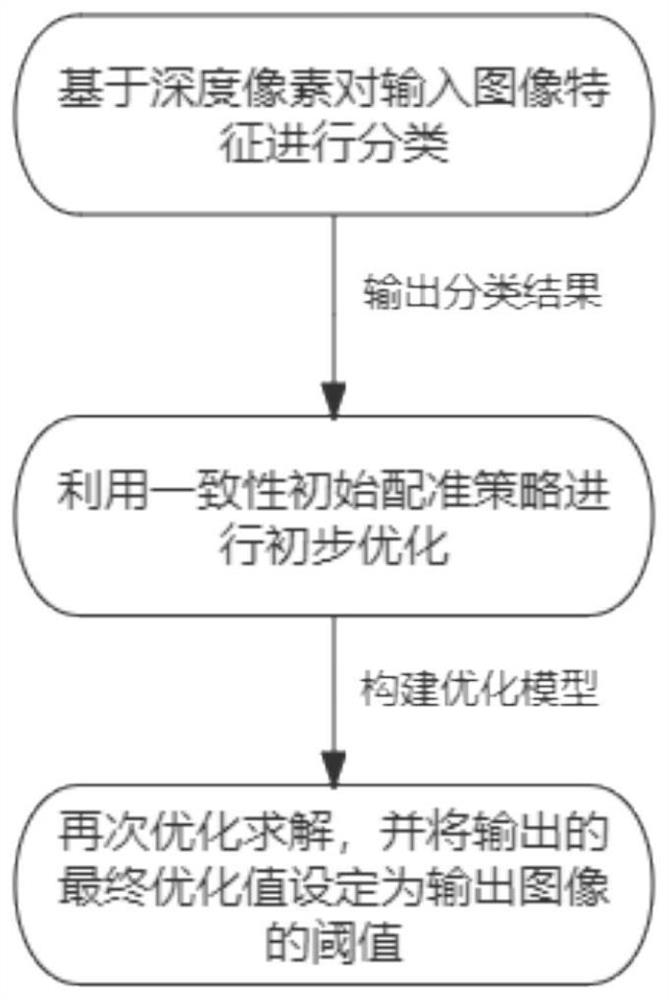
[0052] refer to Figure 1 to Figure 4 , is an implementation provided by an embodiment of the present invention, which specifically includes:

[0053] S1: Classify input image features based on depth pixels, and output classification results. It should be noted that the categories include:

[0054] For the test pixels, the pixel pair composed of the center pixel and each surrounding pixel is classified by the trained CNN, and the final label is determined by a voting strategy.

[0055] Specifically, it also includes:

[0056] Image grayscale (see the image as a three-dimensional image of x, y, z (grayscale));

[0057] Gamma correction method is used to standardize the color space of the input image (normalization); the purpose is to adjust the contrast of the image, reduce the influence of local shadows and illumination changes in the image, and suppress the interference of noise;

[0058] Calculate the gradient of each pixel of the image (including size and orientation); ...

Embodiment 2

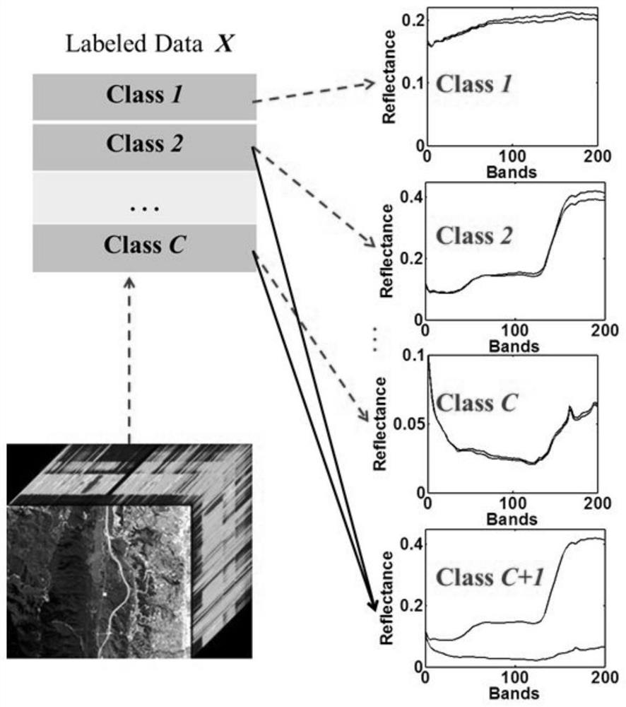
[0088] Further, existing image feature learning methods aim to automatically learn data-adaptive image representations from raw pixel image data. However, existing techniques are poor in extracting and organizing discriminative information from data, and most learning frameworks use unsupervised methods. The method does not consider the information of the class label, so in this embodiment, it is proposed to encode the shareable information in the existing class group, and the discriminant mode has a specific class label in the image feature learning process (that is, the proposed method in Embodiment 1). The classification of the special feature processing) specifically includes:

[0089] Building a Multilayer Feature Learning Framework: Deep Discriminative and Shared Feature Learning.

[0090] Purpose: Hierarchical learning of transform filter banks to transform pixel values ​​of local image patches into features.

[0091] The purpose of each feature learning layer is to le...


PUM


Abstract

The invention discloses a hyperspectral microscopic imaging optimization method suitable for tumor diagnosis, and the method comprises the steps: classifying the features of an input image based on depth pixels, and outputting a classification result; performing preliminary optimization on the classification result by using a consistency initial registration strategy; and constructing an optimization model in combination with a multi-objective optimization strategy, performing optimization solution on the preliminary optimization result again, and setting an output final optimization value as a threshold value of an output image. According to the method, the key feature descriptors are obtained through a special feature classification processing means, the double optimization technology is combined, the accuracy of key information of image processing is guaranteed, on the basis that the key features are reserved, unnecessary complicated calculation is reduced, the imaging speed and accuracy are improved, and ideal help is provided for medical services.

Description

technical field [0001] The invention relates to the technical field of hyperspectral microscopic imaging and image optimization, in particular to a hyperspectral microscopic imaging optimization method suitable for tumor diagnosis. Background technique [0002] At present, in the diagnosis of tumors, especially malignant liver cancer, histopathological diagnosis is still the gold standard. Microscopic endoscopes, electronic endoscopes, ultrasonic endoscopes, etc. have appeared, although they can provide doctors with clearer images, It reduces the suffering of patients, but it is difficult to detect early lesions only through images, which makes early biopsy random. [0003] Spectral imaging technology is a combination of imaging technology and spectral technology, which can simultaneously image the same measured object in a wide continuous spectrum, while detecting the spatial characteristics of the object, each spatial pixel is dispersed into dozens of pixels. To image hun...

Claims


Application Information

Patent Timeline
Patent Type & Authority Applications(China)
IPC IPC(8): G06V10/764G06V10/56G06V10/50G06V20/69
CPCG06V10/764G06V10/56G06V10/50G06V20/69
Inventor 蔡惠明钱露卢露
Owner NANJING NUOYUAN MEDICAL DEVICES CO LTD
Learn More