Semi-supervised learning method for carrying out cell nucleus segmentation on histopathological image

A histopathology, cell nucleus technology, applied in the field of semi-supervised learning, can solve the problems of inconsistent staining operation, difficult to eliminate the difference of image content, difference of image style, etc.

Pending Publication Date: 2022-07-01
CENT SOUTH UNIV
View PDF0 Cites 2 Cited by
  • Summary
  • Abstract
  • Description
  • Claims
  • Application Information

AI Technical Summary

Problems solved by technology

Moreover, this variability in image content is difficult to remove because it already exists when the tissue sample is extracted
In addition, when making histopathological images, there may also be differences in image styles, which come from inconsistent staining operations and different scanning equipment
However, manual labeling of cell nuclei requires professionals and is time-consuming and labor-intensive
However, most current methods that can utilize unlabeled data hardly consider both the segmentation task and the characteristics of histopathological images.

Method used


Image

  • Semi-supervised learning method for carrying out cell nucleus segmentation on histopathological image
  • Semi-supervised learning method for carrying out cell nucleus segmentation on histopathological image
  • Semi-supervised learning method for carrying out cell nucleus segmentation on histopathological image

Examples


Embodiment Construction

[0012] specific implementation

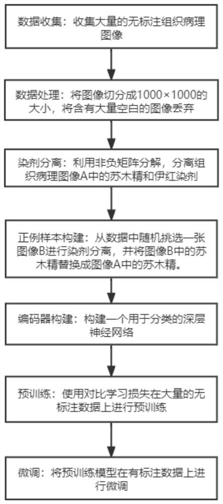
[0013] The specific process of implementing this method is as follows: figure 1 As shown, the semi-supervised learning cell nucleus segmentation method provided by the present invention includes the following steps:

[0014] 1. Obtain a large amount of unlabeled data, that is, histopathological images stained with hematoxylin and eosin (full-field digital sections);

[0015] 2. Process the obtained full-field digital slices, including dividing the large-sized full-field digital slices into smaller images, such as 1000×1000; blank picture;

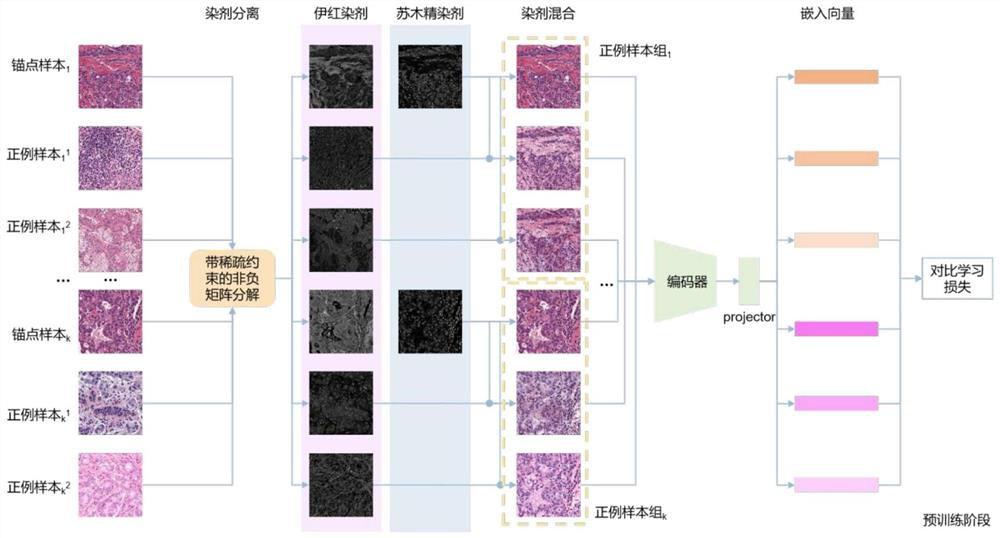
[0016] 3. Using non-negative matrix factorization with sparse constraints to separate unlabeled hematoxylin and eosin-stained histopathological images;

[0017] 4. Select an anchor image, then randomly select two positive sample images for the anchor image, and replace the hematoxylin stain in the positive sample image with the hematoxylin stain in the anchor image, thereby constructing a A set of positi...


PUM


Abstract

The invention discloses a semi-supervised learning method special for performing cell nucleus segmentation on a histopathological image dyed by hematoxylin eosin. According to the cell nucleus segmentation method provided by the invention, according to the characteristics of the histopathological image and cell nucleus segmentation, the two dyes of hematoxylin and eosin in the histopathological image are separated by adopting non-negative matrix factorization with sparse constraint, and then the eosin dye in the histopathological image is replaced by the eosin dye in other histopathological images, so that the segmentation efficiency of the cell nucleus is improved. Therefore, a group of positive example samples can be prepared, and the positive example samples have the same hematoxylin staining agent, so that the positive example samples have interpretable invariance. And inputting the multiple groups of positive example samples into an encoder, and outputting a corresponding embedded representation vector by the encoder. And constraining the model by adopting a contrast learning loss function, so that the model can learn invariance in a positive example sample, namely the hematoxylin staining agent. The hematoxylin stain can stain the cell nucleus and other nucleic acid-rich parts, such as ribosome, so that the hematoxylin stain and the cell nucleus have relatively high correlation. When the model learns the characteristics of the hematoxylin stain, the characteristics accord with the characteristics of a cell nucleus segmentation task, so that the training of the downstream cell nucleus segmentation task is facilitated. As positive example sample construction and pre-training do not need labels, a large amount of unlabeled data can be utilized for training in the mode. And finally, the pre-trained encoder is added into the segmentation model, and fine adjustment is performed on a very small amount of labeled data, so that an effect better than supervised learning on a small amount of samples can be achieved. Therefore, the demand of annotation data is also reduced, and the labor cost is greatly reduced.

Description

technical field [0001] The invention belongs to the field of medical image processing, and relates to a semi-supervised learning method specially used for cell nucleus segmentation of hematoxylin and eosin-stained histopathological images. [0002] technical background [0003] In pathology, the pathological examination is the "gold standard" for the diagnosis of many diseases, including the diagnosis of almost all types of tumors, so millions of biopsies are performed every year. The number, size, shape, density, and nuclear-cytoplasmic ratio of nuclei in tissue sections are all important reference factors for pathologists to diagnose. Medical image segmentation is an important step in medical image analysis, and gradually plays an important role in computer-aided diagnosis and smart medicine. To allow physicians to focus more directly on anatomical and pathological structures of interest when viewing medical images, it is a good option to use medical image segmentation too...

Claims


Application Information

Patent Timeline
Patent Type & Authority Applications(China)
IPC IPC(8): G06T7/00G06T7/11G06N3/04G06N3/08
CPCG06T7/0012G06T7/11G06N3/08G06T2207/20021G06T2207/20081G06T2207/20084G06T2207/30024G06N3/045
Inventor 钟玄同陈先来安莹
Owner CENT SOUTH UNIV
Why Eureka
Try Eureka