
Pathological image classification device and method and use method of device

A pathological image and classification device technology, applied in the field of image recognition and deep learning, can solve the problems of lower classification accuracy and reliability, small pathological image data set, over-fitting phenomenon, etc., to improve the performance of pathological image classification, The effect of reducing the amount of calculation and improving the prediction accuracy

Pending Publication Date: 2022-05-24
BEIJING BOCO COMM TECH
View PDF0 Cites 2 Cited by
  • Summary
  • Abstract
  • Description
  • Claims
  • Application Information

AI Technical Summary

Problems solved by technology

However, because the ResNet convolutional neural network is designed for natural images, in order to obtain more accurate image recognition results, the number of layers required is generally deep, and the calculation is time-consuming; on the other hand, there are many model parameters, and a large amount of training data is required to train the parameters. , in order to obtain higher accuracy
However, due to the small size of the pathological image data set, the above model is directly used in the computer-aided diagnosis system to automatically classify pathological images, which is prone to overfitting and reduces the accuracy and reliability of classification.

Method used


Image

  • Pathological image classification device and method and use method of device
  • Pathological image classification device and method and use method of device
  • Pathological image classification device and method and use method of device

Examples


Embodiment 4

[0145] In order to describe in detail the working principle and use method of a pathological image classification device for the training set and the verification set, the fourth embodiment of the present invention is given, which includes the following steps:

[0146] After selecting a group of morphological digital slices that have been correctly classified, the annotations already contain the original regional classification label and the original pathological classification label, and divided into training set, validation set, and test set according to a certain proportion. Taking the training set and the validation set as the target data of the device, after training, the optimal device parameters are determined.

[0147] Step S401, the image data set generation unit samples the morphological digital slices of the training set to generate a small block image data set 1;

[0148] The image data set generation unit samples the morphological digital slices of the training se...

Embodiment 5

[0165] In order to describe in detail the working principle and use method of a pathological image classification device for the test set, the fifth embodiment of the present invention is given, which includes the following steps:

[0166] After selecting a group of morphological digital slices that have been correctly classified and have included the original regional classification label and the original pathological classification label in the annotation, they are divided into training set, verification set and test set according to a certain proportion. Taking the test set as the target data of the device, the obtained labels can be compared with the original labels to evaluate the performance of the device.

[0167] Step S51, the image data set generation unit samples the morphological digital slices of the test set, and generates a small block image data set 1 of the test set;

[0168] The image data set generation unit samples the morphological digital slices of the tes...


PUM


Abstract

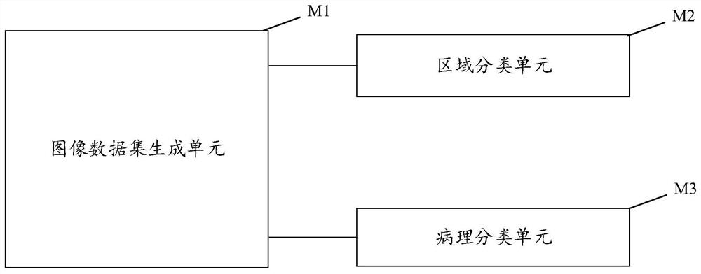
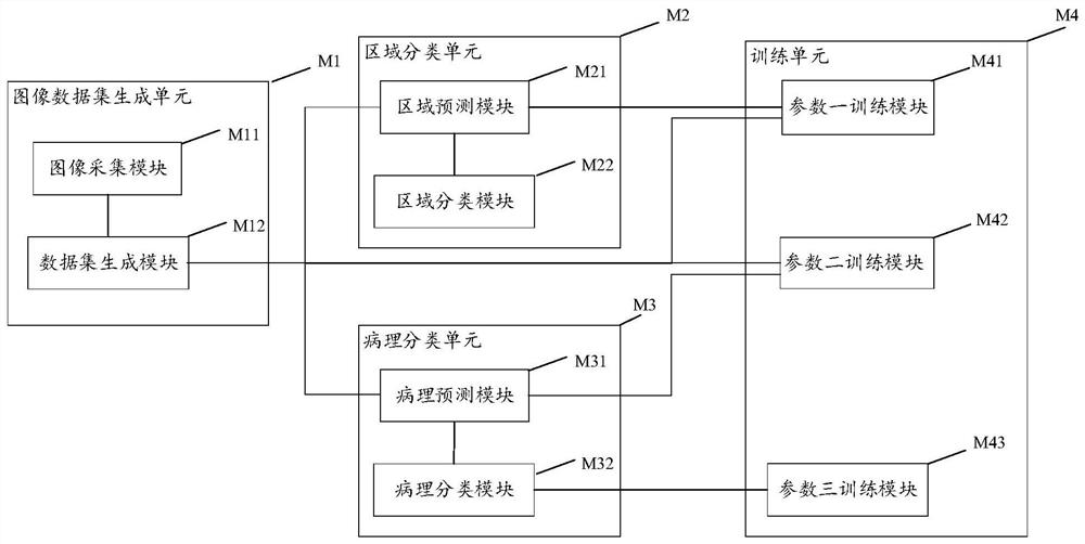
The invention discloses a pathological image classification device, and the device comprises an image data set generation unit which is used for carrying out the sampling of morphological digital slices, and generating a first small image data set; the sampling module is also used for sampling the morphological digital slices and generating a small block image data set II in combination with a set data set rule; the region classification unit is used for calculating prediction region classification labels of the small-block image data in the small-block image data set I; combining a predetermined region classification rule, and synthesizing the small images into regions on the morphological digital slices; the pathology classification unit is used for calculating a predicted pathology classification label of small block abnormal image data in the small block image data set II; and calculating predicted pathological classification labels of the small abnormal image data blocks in the same abnormal area to obtain pathological classification labels of the abnormal areas. The invention further discloses a pathological image classification method. The invention discloses a use method of a pathological image classification device. According to the invention, rapid and accurate pathological image classification can be realized.

Description

technical field [0001] The present invention relates to the field of image recognition and deep learning, and more particularly, to a pathological image classification technology. Background technique [0002] Pathological images are the gold standard for the final diagnosis of cancer. However, the current classification of pathological images based on doctors' manual work is not only time-consuming and labor-intensive, but also the diagnosis results are easily affected by subjective human factors such as doctor's experience and level. The introduction of computer-aided diagnosis systems can not only improve the diagnosis efficiency, but also assist Provide more objective and accurate diagnostic results. In recent years, some convolutional neural network models have been used in computer-aided diagnosis systems to automatically classify pathological images, among which the ResNet convolutional neural network is the most widely used. However, since the ResNet convolutional ...

Claims


Application Information

Patent Timeline
Patent Type & Authority Applications(China)
IPC IPC(8): G06V10/764G06V10/82G06K9/62G16H30/20G06N3/04G06N3/08
CPCG16H30/20G06N3/08G06N3/045G06F18/241
Inventor 魏湘国
Owner BEIJING BOCO COMM TECH
Learn More